Chapter 17
Antihypertensives are drugs that lower blood pressure. Most often they are used for arterial hypertension, i.e. with high blood pressure. Therefore, this group of substances is also called antihypertensive agents.
Arterial hypertension is a symptom of many diseases. There are primary arterial hypertension, or hypertension (essential hypertension), as well as secondary (symptomatic) hypertension, for example, arterial hypertension with glomerulonephritis and nephrotic syndrome (renal hypertension), with narrowing of the renal arteries (renovascular hypertension), pheochromocytoma, hyperaldosteronism, etc.
In all cases, seek to cure the underlying disease. But even if this fails, arterial hypertension should be eliminated, since arterial hypertension contributes to the development of atherosclerosis, angina pectoris, myocardial infarction, heart failure, visual impairment, and impaired renal function. A sharp increase in blood pressure - a hypertensive crisis can lead to bleeding in the brain (hemorrhagic stroke).
In different diseases, the causes of arterial hypertension are different. IN initial stage arterial hypertension is associated with an increase in the tone of the sympathetic nervous system, which leads to an increase in cardiac output and narrowing blood vessels. In this case, blood pressure is effectively reduced by substances that reduce the influence of the sympathetic nervous system (hypotensive agents of central action, adrenoblockers).
In kidney diseases, in the late stages of hypertension, an increase in blood pressure is associated with activation of the renin-angiotensin system. The resulting angiotensin II constricts blood vessels, stimulates the sympathetic system, increases the release of aldosterone, which increases the reabsorption of Na + ions in the renal tubules and thus retains sodium in the body. Drugs that reduce the activity of the renin-angiotensin system should be prescribed.
In pheochromocytoma (a tumor of the adrenal medulla), the adrenaline and norepinephrine secreted by the tumor stimulate the heart, constrict the blood vessels. The pheochromocytoma is removed surgically, but before the operation, during the operation, or, if the operation is not possible, lower the blood pressure with the help of wasp-adrenergic blockers.
common cause arterial hypertension may be a delay in the body of sodium due to excessive consumption of salt and insufficiency of natriuretic factors. An increased content of Na + in the smooth muscles of blood vessels leads to vasoconstriction (the function of the Na + / Ca 2+ exchanger is disturbed: the entry of Na + and the release of Ca 2+ decrease; the level of Ca 2+ in the cytoplasm of smooth muscles increases). As a result, blood pressure rises. Therefore, in arterial hypertension, diuretics are often used that can remove excess sodium from the body.
In arterial hypertension of any genesis, myotropic vasodilators have an antihypertensive effect.
It is believed that in patients with arterial hypertension, antihypertensive drugs should be used systematically, preventing an increase in blood pressure. For this, it is advisable to prescribe long-acting antihypertensive drugs. Most often, drugs are used that act 24 hours and can be administered once a day (atenolol, amlodipine, enalapril, losartan, moxonidine).
In practical medicine, among antihypertensive drugs, diuretics, β-blockers, calcium channel blockers, α-blockers, ACE inhibitors, and AT 1 receptor blockers are most often used.
To stop hypertensive crises, diazoxide, clonidine, azamethonium, labetalol, sodium nitroprusside, nitroglycerin are administered intravenously. In non-severe hypertensive crises, captopril and clonidine are prescribed sublingually.
Classification of antihypertensive drugs
I. Drugs that reduce the influence of the sympathetic nervous system (neurotropic antihypertensive drugs):
1) means of central action,
2) means blocking sympathetic innervation.
P. Myotropic vasodilators:
1) donors N0,
2) potassium channel activators,
3) drugs with an unknown mechanism of action.
III. Calcium channel blockers.
IV. Means that reduce the effects of the renin-angiotensin system:
1) drugs that disrupt the formation of angiotensin II (drugs that reduce renin secretion, ACE inhibitors, vasopeptidase inhibitors),
2) blockers of AT 1 receptors.
V. Diuretics.
Drugs that reduce the effects of the sympathetic nervous system
(neurotropic antihypertensive drugs)
The higher centers of the sympathetic nervous system are located in the hypothalamus. From here, excitation is transmitted to the center of the sympathetic nervous system, located in the rostroventrolateral region of the medulla oblongata (RVLM - rostro-ventrolateral medulla), traditionally called the vasomotor center. From this center, impulses are transmitted to the sympathetic centers of the spinal cord and further along the sympathetic innervation to the heart and blood vessels. Activation of this center leads to an increase in the frequency and strength of heart contractions (increase in cardiac output) and to an increase in the tone of blood vessels - blood pressure rises.
It is possible to reduce blood pressure by inhibiting the centers of the sympathetic nervous system or by blocking the sympathetic innervation. In accordance with this, neurotropic antihypertensive drugs are divided into central and peripheral agents.
TO centrally acting antihypertensives include clonidine, moxonidine, guanfacine, methyldopa.
Clonidine (clophelin, hemiton) - a 2 -adrenomimetic, stimulates a 2A -adrenergic receptors in the center of the baroreceptor reflex in the medulla oblongata (nuclei of the solitary tract). In this case, the centers of the vagus (nucleus ambiguus) and inhibitory neurons are excited, which have a depressing effect on the RVLM (vasomotor center). In addition, the inhibitory effect of clonidine on RVLM is due to the fact that clonidine stimulates I 1 -receptors (imidazoline receptors).
As a result, the inhibitory effect of the vagus on the heart increases and the stimulating effect of sympathetic innervation on the heart and blood vessels decreases. As a result, cardiac output and the tone of blood vessels (arterial and venous) decrease - blood pressure decreases.
In part, the hypotensive effect of clonidine is associated with the activation of presynaptic a 2 -adrenergic receptors at the ends of sympathetic adrenergic fibers - the release of norepinephrine decreases.
At higher doses, clonidine stimulates extrasynaptic a 2 B -adrenergic receptors of smooth muscles of blood vessels (Fig. 45) and, with rapid intravenous administration, can cause short-term vasoconstriction and an increase in blood pressure (therefore, intravenous clonidine is administered slowly, over 5-7 minutes).
In connection with the activation of a 2 -adrenergic receptors of the central nervous system, clonidine has a pronounced sedative effect, potentiates the action of ethanol, and exhibits analgesic properties.
Clonidine is a highly active antihypertensive agent (therapeutic dose when administered orally 0.000075 g); acts for about 12 hours. However, with systematic use, it can cause a subjectively unpleasant sedative effect (absent-mindedness, inability to concentrate), depression, decreased tolerance to alcohol, bradycardia, dry eyes, xerostomia (dry mouth), constipation, impotence. With a sharp cessation of taking the drug, a pronounced withdrawal syndrome develops: after 18-25 hours, blood pressure rises, a hypertensive crisis is possible. β-Adrenergic blockers increase the clonidine withdrawal syndrome, so these drugs are not prescribed together.
Clonidine is mainly used to quickly lower blood pressure in hypertensive crises. In this case, clonidine is administered intravenously over 5-7 minutes; with rapid administration, an increase in blood pressure is possible due to stimulation of a 2 -adrenergic receptors of blood vessels.
Clonidine solutions in the form of eye drops are used in the treatment of glaucoma (reduces the production of intraocular fluid).
Moxonidine(cint) stimulates imidazoline 1 1 receptors in the medulla oblongata and, to a lesser extent, a 2 adrenoreceptors. As a result, the activity of the vasomotor center decreases, cardiac output and the tone of blood vessels decrease - blood pressure decreases.
The drug is prescribed orally for the systematic treatment of arterial hypertension 1 time per day. Unlike clonidine, when using moxonidine, sedation, dry mouth, constipation, and withdrawal syndrome are less pronounced.
Guanfacine(Estulik) similarly to clonidine stimulates central a 2 -adrenergic receptors. Unlike clonidine, it does not affect 1 1 receptors. The duration of the hypotensive effect is about 24 hours. Assign inside for the systematic treatment of arterial hypertension. The withdrawal syndrome is less pronounced than that of clonidine. 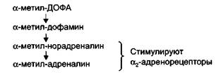
Methyldopa(dopegit, aldomet) according to the chemical structure - a-methyl-DOPA. The drug is prescribed inside. In the body, methyldopa is converted to methylnorepinephrine, and then to methyladrenaline, which stimulate the a 2 -adrenergic receptors of the center of the baroreceptor reflex.
Metabolism of methyldopa
The hypotensive effect of the drug develops after 3-4 hours and lasts about 24 hours.
Side effects of methyldopa: dizziness, sedation, depression, nasal congestion, bradycardia, dry mouth, nausea, constipation, liver dysfunction, leukopenia, thrombocytopenia. In connection with the blocking effect of a-methyl-dopamine on dopaminergic transmission, the following are possible: parkinsonism, increased production of prolactin, galactorrhea, amenorrhea, impotence (prolactin inhibits the production of gonadotropic hormones). With a sharp discontinuation of the drug, the withdrawal syndrome manifests itself after 48 hours.
Drugs that block peripheral sympathetic innervation.
To reduce blood pressure, sympathetic innervation can be blocked at the level of: 1) sympathetic ganglia, 2) endings of postganglionic sympathetic (adrenergic) fibers, 3) adrenoreceptors of the heart and blood vessels. Accordingly, ganglioblockers, sympatholytics, adrenoblockers are used.
Ganglioblockers - hexamethonium benzosulfonate(benzo-hexonium), azamethonium(pentamine), trimetaphan(arfonad) block the transmission of excitation in the sympathetic ganglia (block N N -xo-linoreceptors of ganglionic neurons), block N N -cholinergic receptors of the chromaffin cells of the adrenal medulla and reduce the release of adrenaline and norepinephrine. Thus, ganglion blockers reduce the stimulating effect of sympathetic innervation and catecholamines on the heart and blood vessels. There is a weakening of the contractions of the heart and the expansion of arterial and venous vessels - arterial and venous pressure decreases. At the same time, ganglion blockers block the parasympathetic ganglia; thus eliminate the inhibitory effect of the vagus nerves on the heart and usually cause tachycardia.
Ganglioblockers are not very suitable for systematic use due to side effects (severe orthostatic hypotension, disturbance of accommodation, dry mouth, tachycardia; intestinal atony and Bladder, sexual dysfunction).
Hexamethonium and azamethonium act for 2.5-3 hours; administered intramuscularly or under the skin in hypertensive crises. Azamethonium is also administered intravenously slowly in 20 ml of isotonic sodium chloride solution in case of a hypertensive crisis, swelling of the brain, lungs against the background of high blood pressure, with spasms of peripheral vessels, with intestinal, hepatic or renal colic.
Trimetafan acts 10-15 minutes; is administered in solutions intravenously by drip for controlled hypotension during surgical operations.
Sympatholytics- reserpine, guanethidine(octadin) reduce the release of norepinephrine from the endings of sympathetic fibers and thus reduce the stimulating effect of sympathetic innervation on the heart and blood vessels - arterial and venous pressure decreases. Reserpine reduces the content of norepinephrine, dopamine and serotonin in the central nervous system, as well as the content of adrenaline and norepinephrine in the adrenal glands. Guanethidine does not penetrate the blood-brain barrier and does not change the content of catecholamines in the adrenal glands.
Both drugs differ in the duration of action: after the systematic administration is stopped, the hypotensive effect can persist for up to 2 weeks. Guanethidine is much more effective than reserpine, but due to severe side effects, it is rarely used.
In connection with the selective blockade of sympathetic innervation, the influences of the parasympathetic nervous system predominate. Therefore, when using sympatholytics, the following are possible: bradycardia, increased secretion of HC1 (contraindicated in peptic ulcer), diarrhea. Guanethidine causes significant orthostatic hypotension (associated with a decrease in venous pressure); when using reserpine, orthostatic hypotension is not very pronounced. Reserpine reduces the level of monoamines in the central nervous system, can cause sedation, depression.
A -Ldrenoblockers reduce the ability to stimulate the effect of sympathetic innervation on blood vessels (arteries and veins). In connection with the expansion of blood vessels, arterial and venous pressure decreases; heart contractions reflexively increase.
a 1 - Adrenoblockers - prazosin(minipress), doxazosin, terazosin administered orally for the systematic treatment of arterial hypertension. Prazosin acts 10-12 hours, doxazosin and terazosin - 18-24 hours.
Side effects of a 1 -blockers: dizziness, nasal congestion, moderate orthostatic hypotension, tachycardia, frequent urination.
a 1 a 2 - Adrenoblocker phentolamine used for pheochromocytoma before surgery and during surgery to remove pheochromocytoma, as well as in cases where surgery is not possible.
β -Adrenoblockers- one of the most commonly used groups of antihypertensive drugs. With systematic use, they cause a persistent hypotensive effect, prevent sharp rises in blood pressure, practically do not cause orthostatic hypotension, and, in addition to hypotensive properties, have antianginal and antiarrhythmic properties.
β-blockers weaken and slow down the contractions of the heart - systolic blood pressure decreases. At the same time, β-blockers constrict blood vessels (block β 2 -adrenergic receptors). Therefore, with a single use of β-blockers, mean arterial pressure usually decreases slightly (with isolated systolic hypertension, blood pressure may decrease after a single use of β-blockers).
However, if p-blockers are used systematically, then after 1-2 weeks, vasoconstriction is replaced by their expansion - blood pressure decreases. Vasodilation is explained by the fact that with the systematic use of β-blockers, due to a decrease in cardiac output, the baroreceptor depressor reflex is restored, which is weakened in arterial hypertension. In addition, vasodilation is facilitated by a decrease in renin secretion by juxtaglomerular cells of the kidneys (block of β 1 -adrenergic receptors), as well as blockade of presynaptic β 2 -adrenergic receptors at the endings of adrenergic fibers and a decrease in the release of norepinephrine.
For the systematic treatment of arterial hypertension, long-acting β 1 -adrenergic blockers are more often used - atenolol(tenormin; lasts about 24 hours), betaxolol(valid up to 36 hours).
Side effects of β-adrenergic blockers: bradycardia, heart failure, atrioventricular conduction difficulty, decreased plasma HDL levels, increased bronchial and peripheral vascular tone (less pronounced in β 1-blockers), increased action of hypoglycemic agents, decreased physical activity.
a 2 β -Adrenoblockers - labetalol(transat), carvedilol(dilatrend) reduce cardiac output (block of p-adrenergic receptors) and reduce the tone of peripheral vessels (block of a-adrenergic receptors). The drugs are used orally for the systematic treatment of arterial hypertension. Labetalol is also administered intravenously in hypertensive crises.
Carvedilol is also used in chronic heart failure.
| Sympathetic department | Parasympathetic department |
| 1. Accelerates the rhythm, increases the strength of heart contractions 2. Expands the coronary vessels of the heart 3. Constricts most of the blood vessels (internal organs, skin and mucous membranes) 4. Expands the vessels of the brain and skeletal muscles 5. Constricts the veins 6. Does not affect 7. Increases blood pressure and speed of blood movement 8. Expands the bronchi, increases respiration (pulmonary ventilation) 9. Slows down the secretion of juice, tone and peristalsis in the digestive organs (digestion inhibition) 10. Contracts the spleen, expels blood from it 11. Constricts the kidney vessels, reduces urine formation (diuresis) , slows down the kidneys 12. Closes the sphincter, delays urination 13. Stimulates, increases sweating 14. Expands the pupils 15. Increases energy metabolism (dissimilation), increases the release of energy; slows down assimilation, synthesis 16. Breakdown of glycogen and liver fat to glucose and fatty acids, mobilization of organic depots 17. Relaxes the bile ducts 18. Contracts the muscles that raise the hair 19. Provides reactions of “fight or flight” activity 20 Weakening of sexual activity | 1. Slows down the rhythm, reduces the force of heart contractions 2. Narrows the coronary vessels of the heart 3. Does not affect the diameter of the vessels (does not innervate) - 4. Narrows the vessels of the brain and skeletal muscles - 5. Does not affect 6. Expands the vessels of the genital organs 7. Reduces blood pressure and blood velocity 8. Narrows the bronchi, slows down breathing (pulmonary ventilation) 9. Increased secretion of juice, tone and peristalsis in the digestive organs (increased digestion) 10. Does not affect 11. Does not affect 12. Increases the tone of the bladder, relaxes the sphincter, promotes bladder emptying, 13. Weakens 14. Constricts pupils 15. Lowers the level of energy metabolism, reduces energy release, enhances assimilation, synthesis of substances 16. Glycogen formation, fat synthesis, accumulation of reserve organic substances 17. Bile ducts are reduced 18. Does not affect 19 Providing reactions of "rest and recuperation" 20. Increased sexual activity. |
The central regulation of the functions of the autonomic nervous system is carried out cerebral cortex through the hypothalamus and brainstem (mainly through the spinal cord)
Coordination of motor (motor) and vegetative (metabolism, blood circulation, respiration, digestion, excretion, etc.) functions is carried out by the limbic system and the frontal lobes of the cerebral cortex
End of work -
This topic belongs to:
Essence of life
Living matter qualitatively differs from non-living matter in its enormous complexity and high structural and functional orderliness... Living and non-living matter are similar at the elementary chemical level, i.e.... Chemical compounds of cell matter...
If you need additional material on this topic, or you did not find what you were looking for, we recommend using the search in our database of works:
What will we do with the received material:
If this material turned out to be useful for you, you can save it to your page on social networks:
| tweet |
All topics in this section:
III. Mutation process and reserve of hereditary variability
In the gene pool of populations, a continuous mutation process occurs under the influence of mutagenic factors Recessive alleles mutate more often (encode less resistant to the action of mutagenic fa
VI. Allele and genotype frequencies (population genetic structure)
The genetic structure of a population is the ratio of the frequencies of alleles (A and a) and genotypes (AA, Aa, aa) in the gene pool of the population Allele frequency
Cytoplasmic inheritance
There are data that are inexplicable from the point of view of the chromosome theory of heredity by A. Weisman and T. Morgan (i.e., exclusively nuclear localization of genes) The cytoplasm is involved in re
Plasmogenes of mitochondria
One myotochondria contains 4-5 circular DNA molecules about 15,000 base pairs long Contains genes for: - synthesis of t RNA, p RNA and ribosome proteins, some aero enzymes
Plasmids
Plasmids are very short, autonomously replicating circular fragments of the bacterial DNA molecule that provide non-chromosomal transmission of hereditary information.
VARIABILITY
Variability is a common property of all organisms to acquire structural and functional differences from their ancestors.
Mutational variability
Mutations - qualitative or quantitative DNA of body cells, leading to changes in their genetic apparatus (genotype) Mutation theory of creation
Causes of Mutations
Mutagenic factors (mutagens) - substances and influences capable of inducing a mutational effect (any factors of the external and internal environment that can
Mutation frequency
· The frequency of mutation of individual genes varies widely and depends on the state of the organism and the stage of ontogeny (usually increases with age). On average, each gene mutates once every 40,000 years.
Gene mutations (point, true)
The reason is a change in the chemical structure of the gene (violation of the nucleotide sequence in DNA: * gene inserts of a pair or several nucleotides
Chromosomal mutations (chromosomal rearrangements, aberrations)
Causes - are caused by significant changes in the structure of chromosomes (redistribution of the hereditary material of chromosomes) In all cases, they arise as a result of ra
Polyploidy
Polyploidy - a multiple increase in the number of chromosomes in a cell (the haploid set of chromosomes -n is repeated not 2 times, but many times - up to 10 -1
The meaning of polyploidy
1. Polyploidy in plants is characterized by an increase in the size of cells, vegetative and generative organs - leaves, stems, flowers, fruits, root crops, etc. , y
Aneuploidy (heteroploidy)
Aneuploidy (heteroploidy) - a change in the number of individual chromosomes that is not a multiple of the haploid set (in this case, one or more chromosomes from a homologous pair are normal
Somatic mutations
Somatic mutations - mutations that occur in the somatic cells of the body Distinguish between gene, chromosomal and genomic somatic mutations
The law of homologous series in hereditary variability
· Discovered by N. I. Vavilov on the basis of the study of wild and cultivated flora of five continents 5. The mutation process in genetically related species and genera proceeds in parallel, in
Combination variability
Combinative variability - variability resulting from the regular recombination of alleles in the genotypes of offspring, due to sexual reproduction
Phenotypic variability (modification or non-hereditary)
Modification variability - evolutionarily fixed adaptive reactions of an organism to a change in the external environment without changing the genotype
The value of modification variability
1. most modifications have an adaptive value and contribute to the adaptation of the body to a change in the external environment 2. can cause negative changes - morphoses
Statistical patterns of modification variability
· Modifications of a single trait or property, measured quantitatively, form a continuous series (variation series); it cannot be built according to an unmeasurable feature or a feature that exists
Variation curve of the distribution of modifications in the variation series
V - trait variants P - frequency of occurrence of trait variants Mo - mode, or most
Differences in the manifestation of mutations and modifications
Mutational (genotypic) variability Modification (phenotypic) variability 1. Associated with changes in the geno- and karyotype
Features of a person as an object of genetic research
1. It is impossible to purposefully select parental pairs and experimental marriages (impossibility of experimental crossing) 2. Slow generational change, which occurs on average after
Methods for studying human genetics
Genealogical method · The method is based on the compilation and analysis of genealogies (introduced into science at the end of the 19th century by F. Galton); the essence of the method is to trace us
twin method
The method consists in studying the patterns of inheritance of traits in single and dizygotic twins (the frequency of birth of twins is one case per 84 newborns)
Cytogenetic method
Consists of a visual study of mitotic metaphase chromosomes under a microscope Based on the method of differential staining of chromosomes (T. Kasperson,
Dermatoglyphics method
Based on the study of the relief of the skin on the fingers, palms and plantar surfaces of the feet (there are epidermal protrusions - ridges that form complex patterns), this trait is inherited
Population-statistical method
Based on the statistical (mathematical) processing of data on inheritance in large population groups (populations - groups that differ in nationality, religion, race, profession)
Somatic cell hybridization method
Based on the reproduction of somatic cells of organs and tissues outside the body in sterile nutrient media (cells are most often obtained from the skin, bone marrow, blood, embryos, tumors) and
Modeling method
· The theoretical basis of biological modeling in genetics is given by the law of homological series of hereditary variability by N.I. Vavilova For modeling, certain
Genetics and medicine (medical genetics)
Studying the causes, diagnostic signs, possibilities of rehabilitation and prevention of human hereditary diseases (monitoring of genetic abnormalities)
Chromosomal diseases
The reason is a change in the number (genomic mutations) or structure of chromosomes (chromosomal mutations) of the karyotype of the germ cells of the parents (anomalies can occur at different
Polysomy on sex chromosomes
Trisomy - X (Triplo X syndrome); Karyotype (47, XXX) Known in women; syndrome frequency 1: 700 (0.1%) N
Hereditary diseases of gene mutations
Cause - gene (point) mutations (changes in the nucleotide composition of a gene - insertions, substitutions, dropouts, transfers of one or more nucleotides; the exact number of genes in a person is unknown
Diseases controlled by genes located on the X or Y chromosome
Hemophilia - blood incoagulability Hypophosphatemia - loss of phosphorus and lack of calcium by the body, softening of the bones Muscular dystrophy - structural disorders
Genotypic level of prevention
1. Search and application of antimutagenic protective substances Antimutagens (protectors) are compounds that neutralize a mutagen before it reacts with a DNA molecule or remove it
Treatment of hereditary diseases
1. Symptomatic and pathogenetic - impact on the symptoms of the disease (the genetic defect is preserved and transmitted to offspring) n dieter
Gene Interaction
Heredity - a set of genetic mechanisms that ensure the preservation and transmission of the structural and functional organization of a species in a number of generations from ancestors
Interaction of allelic genes (one allelic pair)
There are five types of allelic interactions: 1. Complete dominance 2. Incomplete dominance 3. Overdominance 4. Codominance
complementarity
Complementarity - the phenomenon of the interaction of several non-allelic dominant genes, leading to the emergence of a new trait that is absent in both parents
Polymerism
Polymeria - the interaction of non-allelic genes, in which the development of one trait occurs only under the action of several non-allelic dominant genes (polygene
Pleiotropy (multiple gene action)
Pleiotropy - the phenomenon of the influence of one gene on the development of several traits The reason for the pleiotropic influence of a gene is in the action of the primary product of this
Selection basics
Selection (lat. selektio - selection) - science and industry of agricultural. production, developing the theory and methods of creating new and improving existing plant varieties, animal breeds
Domestication as the first stage of selection
Cultivated plants and domestic animals are descended from wild ancestors; this process is called domestication or domestication The driving force behind domestication is the suit
Centers of origin and diversity of cultivated plants (according to N. I. Vavilov)
Center name Geographical location Homeland of cultivated plants
Artificial selection (selection of parent pairs)
Two types of artificial selection are known: mass and individual
Hybridization (crossing)
Allows you to combine certain hereditary traits in one organism, as well as get rid of undesirable properties In breeding, various crossing systems are used &n
Inbreeding (inbreeding)
Inbreeding is the crossing of individuals with a close degree of kinship: brother - sister, parents - offspring (in plants, the closest form of inbreeding occurs when self-breeding
Outbreeding (outbreeding)
When crossing unrelated individuals, harmful recessive mutations that are in the homozygous state become heterozygous and do not adversely affect the viability of the organism
heterosis
Heterosis (hybrid strength) is a phenomenon of a sharp increase in the viability and productivity of first-generation hybrids during unrelated crossing (interbreeding).
Induced (artificial) mutagenesis
The frequency with the spectrum of mutations increases dramatically when exposed to mutagens (ionizing radiation, chemicals, extreme environmental conditions, etc.)
Interline hybridization in plants
It consists in crossing pure (inbred) lines obtained as a result of long-term forced self-pollination of cross-pollinated plants in order to obtain maximum
Vegetative propagation of somatic mutations in plants
The method is based on the isolation and selection of useful somatic mutations for economic traits in the best old varieties (possible only in plant breeding)
Methods of breeding and genetic work by I. V. Michurina
1. Systematically distant hybridization
Polyploidy
Polyploidy - the phenomenon of a multiple of the main number (n) of an increase in the number of chromosomes in the somatic cells of the body (the mechanism for the formation of polyploids and
Cell engineering
Cultivation of individual cells or tissues on artificial sterile nutrient media containing amino acids, hormones, mineral salts and other nutritional components (
Chromosomal engineering
The method is based on the possibility of replacing or adding new individual chromosomes in plants It is possible to decrease or increase the number of chromosomes in any homologous pair - aneuploidy
Animal breeding
Has a number of features in comparison with plant breeding, which objectively make it difficult to carry out 1. Only sexual reproduction is characteristic (lack of vegetative
domestication
It began about 10 - 5 thousand years ago in the Neolithic era (it weakened the effect of stabilizing natural selection, which led to an increase in hereditary variability and an increase in the selection efficiency
Crossing (hybridization)
There are two methods of crossing: related (inbreeding) and unrelated (outbreeding) When selecting a pair, the pedigrees of each manufacturer are taken into account (stud books, learn
Outbreeding (outbreeding)
Can be intrabreeding and interbreeding, interspecific or intergeneric (systematically distant hybridization) Accompanied by the effect of heterosis of F1 hybrids
Checking the breeding qualities of producers by offspring
There are economic traits that appear only in females (egg production, milk production) Males are involved in the formation of these traits in daughters (it is necessary to check males for c
Selection of microorganisms
Microorganisms (prokaryotes - bacteria, blue-green algae; eukaryotes - unicellular algae, fungi, protozoa) - are widely used in industry, agriculture, medicine
Stages of selection of microorganisms
I. The search for natural strains capable of synthesizing the products necessary for a person II. The isolation of a pure natural strain (occurs in the process of repeated seeding of
Tasks of biotechnology
1. Obtaining feed and food protein from cheap natural raw materials and industrial waste (the basis for solving the food problem) 2. Obtaining a sufficient amount
Products of microbiological synthesis
q Feed and food protein q Enzymes (widely used in food, alcohol, brewing, winemaking, meat, fish, leather, textile, etc.)
Stages of the technological process of microbiological synthesis
Stage I - obtaining a pure culture of microorganisms containing only organisms of one species or strain Each species is stored in a separate test tube and goes to production and
Genetic (genetic) engineering
Genetic engineering is a field of molecular biology and biotechnology that deals with the creation and cloning of new genetic structures (recombinant DNA) and organisms with specified characteristics.
Stages of obtaining recombinant (hybrid) DNA molecules
1. Obtaining the original genetic material - the gene encoding the protein (trait) of interest The necessary gene can be obtained in two ways: artificial synthesis or extraction
Achievements in genetic engineering
The introduction of eukaryotic genes into bacteria is used for the microbiological synthesis of biologically active substances, which in nature are synthesized only by cells of higher organisms Synthesis
Problems and prospects of genetic engineering
Study of the molecular basis of hereditary diseases and the development of new methods for their treatment, finding methods for correcting damage to individual genes Increasing the resistance of the organ
Chromosomal engineering in plants
It consists in the possibility of biotechnological replacement of individual chromosomes in plant gametes or the addition of new ones In the cells of each diploid organism there are pairs of homologous chromosomes
Cell and tissue culture method
The method is the cultivation of individual cells, pieces of tissue or organs outside the body under artificial conditions on strictly sterile nutrient media with constant physical and chemical
Clonial micropropagation of plants
Cultivation of plant cells is relatively uncomplicated, the media are simple and cheap, and cell culture is unpretentious The method of plant cell culture is that a single cell or t
Hybridization of somatic cells (somatic hybridization) in plants
Protoplasts of plant cells without rigid cell walls can merge with each other, forming a hybrid cell that has the characteristics of both parents Gives the opportunity to receive
Cellular engineering in animals
Method of hormonal superovulation and embryo transplantation Isolation of dozens of eggs per year from the best cows by the method of hormonal inductive poliovulation (called
Hybridization of somatic cells in animals
Somatic cells contain the entire amount of genetic information Somatic cells for cultivation and subsequent hybridization in humans are obtained from the skin, which
Obtaining monoclonal antibodies
In response to the introduction of an antigen (bacteria, viruses, erythrocytes, etc.), the body produces specific antibodies with the help of B-lymphocytes, which are proteins called imm
Environmental Biotechnology
· Purification of water through the creation of wastewater treatment plants using biological methods q Oxidation of wastewater on biological filters q Utilization of organic and
Bioenergy
Bioenergy is a direction of biotechnology associated with obtaining energy from biomass with the help of microorganisms One of the effective methods for obtaining energy from biome
Bioconversion
Bioconversion is the conversion of substances formed as a result of metabolism into structurally related compounds under the action of microorganisms The goal of bioconversion is
Engineering enzymology
Engineering enzymology is a field of biotechnology that uses enzymes in the production of given substances The central method of engineering enzymology is immobilization
Biogeotechnology
Biogeotechnology - the use of the geochemical activity of microorganisms in the mining industry (ore, oil, coal) With the help of micro
The boundaries of the biosphere
Determined by a complex of factors; the general conditions for the existence of living organisms include: 1. the presence of liquid water 2. the presence of a number of biogenic elements (macro- and microelements
Properties of living matter
1. They contain a huge supply of energy capable of doing work 2. The speed of chemical reactions in living matter is millions of times faster than usual due to the participation of enzymes
Functions of living matter
Performed by living matter in the process of vital activity and biochemical transformations of substances in metabolic reactions 1. Energy - transformation and assimilation by living
Land biomass
Continental part of the biosphere - land occupies 29% (148 million km2) Land heterogeneity is expressed by the presence of latitudinal zonality and altitudinal zonality
soil biomass
Soil - a mixture of decomposed organic and weathered minerals; the mineral composition of the soil includes silica (up to 50%), alumina (up to 25%), oxide of iron, magnesium, potassium, phosphorus
Biomass of the oceans
The area of the World Ocean (Earth's hydrosphere) occupies 72.2% of the entire surface of the Earth Water has special properties, important for the life of organisms - high heat capacity and heat conductivity
Biological (biotic, biogenic, biogeochemical cycle) cycle of substances
The biotic cycle of substances is a continuous, planetary, relatively cyclic, irregular distribution of substances in time and space.
Biogeochemical cycles of individual chemical elements
Biogenic elements circulate in the biosphere, that is, they perform closed biogeochemical cycles that function under the influence of biological (life activity) and geological
nitrogen cycle
The source of N2 is molecular, gaseous, atmospheric nitrogen (it is not absorbed by most living organisms, because it is chemically inert; plants are able to assimilate only associated with ki
The carbon cycle
The main source of carbon is carbon dioxide of the atmosphere and water The carbon cycle is carried out through the processes of photosynthesis and cellular respiration The cycle begins with f
The water cycle
Carried out by solar energy Regulated by living organisms: 1. absorption and evaporation by plants 2. photolysis in the process of photosynthesis (decomposition
Sulfur cycle
Sulfur is a biogenic element of living matter; found in proteins as part of amino acids (up to 2.5%), is part of vitamins, glycosides, coenzymes, is found in vegetable essential oils
Energy flow in the biosphere
Source of energy in the biosphere - continuous electromagnetic radiation of the sun and radioactive energy q 42% of solar energy is reflected from clouds, dust atmosphere and the Earth's surface in
The emergence and evolution of the biosphere
Living matter, and with it the biosphere, appeared on Earth as a result of the emergence of life in the process of chemical evolution about 3.5 billion years ago, which led to the formation of organic substances
Noosphere
The noosphere (literally, the sphere of the mind) is the highest stage in the development of the biosphere, associated with the emergence and formation of civilized humanity in it, when its mind
Signs of the modern noosphere
1. Increasing amount of recoverable materials of the lithosphere - growth in the development of mineral deposits (now it exceeds 100 billion tons per year) 2. Mass consumption
Human influence on the biosphere
The current state of the noosphere is characterized by an ever-increasing prospect of an ecological crisis, many aspects of which are already manifesting themselves in full, creating a real threat to the existence
Energy production
q The construction of hydroelectric power plants and the creation of reservoirs causes flooding of large areas and the resettlement of people, raising the level of groundwater, erosion and waterlogging of the soil, landslides, loss of arable land
Food production. Depletion and pollution of the soil, reduction of the area of fertile soils
q Arable land covers 10% of the Earth's surface (1.2 billion ha) q Cause - overexploitation, imperfection of agricultural production: water and wind erosion and the formation of ravines, in
Reduction of natural biological diversity
q Human economic activity in nature is accompanied by a change in the number of animal and plant species, the extinction of entire taxa, and a decrease in the diversity of living things.
acid rain
q Increased acidity of rains, snow, fogs due to the emission of sulfur and nitrogen oxides from fuel combustion into the atmosphere q Acid precipitation reduces crops, destroys natural vegetation
Ways to solve environmental problems
In the future, a person will exploit the resources of the biosphere on an ever-increasing scale, since this exploitation is an indispensable and main condition for the very existence of h
Sustainable consumption and management of natural resources
q The most complete and comprehensive extraction of all minerals from the fields (due to the imperfection of the extraction technology, only 30-50% of the reserves are extracted from oil fields q Rec
Ecological strategy for the development of agriculture
q Strategic direction - increasing crop yields to feed a growing population without increasing acreage q Increasing crop yields without negative
Properties of living matter
1. The unity of the elemental chemical composition (98% is carbon, hydrogen, oxygen and nitrogen) 2. The unity of the biochemical composition - all living organisms
Hypotheses for the origin of life on Earth
There are two alternative concepts of the possibility of the origin of life on Earth: q abiogenesis - the emergence of living organisms from substances of inorganic nature
Stages of the development of the Earth (chemical prerequisites for the emergence of life)
1. The stellar stage of the Earth's history q The geological history of the Earth began more than 6 years ago. years ago, when the Earth was a red-hot over 1000
III. The emergence of the process of self-reproduction of molecules (biogenic matrix synthesis of biopolymers)
1. Occurred as a result of the interaction of coacervates with nucleic acids 2. All the necessary components of the process of biogenic matrix synthesis: - enzymes - proteins - pr
Prerequisites for the emergence of the evolutionary theory of Ch. Darwin
Socio-economic background 1. In the first half of the XIX century. England has become one of the most economically developed countries in the world with a high level of
· Set out in the book of Ch. Darwin "On the origin of species by natural selection or the preservation of favored breeds in the struggle for life", which was published
Variability
Substantiation of the variability of species To substantiate the position on the variability of living beings, Charles Darwin used common
Correlative (relative) variability
A change in the structure or function of one part of the body causes a coordinated change in the other or others, since the body is an integral system, the individual parts of which are closely interconnected
The main provisions of the evolutionary teachings of Ch. Darwin
1. All kinds of living creatures inhabiting the Earth have never been created by anyone, but arose naturally 2. Having arisen naturally, species slowly and gradually
The development of ideas about the form
Aristotle - used the concept of species when describing animals, which had no scientific content and was used as a logical concept D. Ray
Species criteria (signs of species identification)
Significance of species criteria in science and practice - determination of species belonging of individuals (species identification) I. Morphological - similarity of morphological inheritances
Population types
1. Panmictic - consist of individuals that reproduce sexually, cross-fertilized. 2. Clonial - from individuals that breed only without
mutation process
Spontaneous changes in the hereditary material of germ cells in the form of gene, chromosome and genomic mutations occur constantly throughout the entire period of life under the influence of mutations
Insulation
Isolation - cessation of the flow of genes from population to population (limitation of the exchange of genetic information between populations) The value of isolation as a fa
Primary insulation
Not directly related to the action of natural selection, is a consequence of external factors Leads to a sharp decrease or cessation of migration of individuals from other populations
Environmental isolation
· Arises on the basis of ecological differences in the existence of different populations (different populations occupy different ecological niches) v For example, the trout of Lake Sevan
Secondary isolation (biological, reproductive)
Is of decisive importance in the formation of reproductive isolation Arises as a result of intraspecific differences in organisms Arose as a result of evolution Has two iso
Migrations
Migrations - the movement of individuals (seeds, pollen, spores) and their characteristic alleles between populations, leading to a change in the frequencies of alleles and genotypes in their gene pools
population waves
Population waves ("waves of life") - periodic and non-periodic sharp fluctuations in the number of individuals in a population under the influence of natural causes (S. S.
Significance of population waves
1. Leads to an undirected and abrupt change in the frequencies of alleles and genotypes in the gene pool of populations (random survival of individuals during the wintering period can increase the concentration of this mutation by 1000 r
Gene drift (genetic-automatic processes)
Genetic drift (genetic-automatic processes) - random non-directional, not due to the action of natural selection, change in the frequencies of alleles and genotypes in m
The result of genetic drift (for small populations)
1. Causes the loss (p = 0) or fixation (p = 1) of alleles in the homozygous state in all members of the population, regardless of their adaptive value - homozygotization of individuals
Natural selection is the guiding factor of evolution
Natural selection is the process of preferential (selective, selective) survival and reproduction of the fittest individuals and non-survival or non-reproduction
Struggle for existence Forms of natural selection
Driving selection (Described by C. Darwin, modern teaching developed by D. Simpson, English) Driving selection - selection in
Stabilizing selection
· The theory of stabilizing selection was developed by the Russian acad. I. I. Shmagauzen (1946) Stabilizing selection - selection acting in stable
Other forms of natural selection
Individual selection - selective survival and reproduction of individuals that have an advantage in the struggle for existence and elimination of others
The main features of natural and artificial selection
Natural selection Artificial selection 1. Arose with the emergence of life on Earth (about 3 billion years ago) 1. Arose in the
Common features of natural and artificial selection
1. Initial (elementary) material - individual characteristics of the organism (hereditary changes - mutations) 2. Carried out according to the phenotype 3. Elementary structure - population
The struggle for existence is the most important factor in evolution
The struggle for existence is a complex relationship of an organism with abiotic (physical conditions of life) and biotic (relations with other living organisms) fact
Reproduction intensity
v One roundworm produces 200 thousand eggs per day; the gray rat gives 5 litters per year, 8 rats, which become sexually mature at the age of three months; offspring of one daphnia per summer
Interspecies struggle for existence
Occurs between individuals of populations of different species Less acute than intraspecific, but its intensity increases if different species occupy similar ecological niches and have
Fight against adverse abiotic environmental factors
It is observed in all cases when individuals of the population find themselves in extreme physical conditions (excessive heat, drought, severe winter, excessive humidity, infertile soils, severe
The main discoveries in the field of biology after the creation of STE
1. Discovery of the hierarchical structures of DNA and protein, including the secondary structure of DNA - the double helix and its nucleoprotein nature 2. Deciphering the genetic code (its triplet
Signs of the organs of the endocrine system
1. They are relatively small in size (fractions or a few grams) 2. Anatomically unrelated 3. Synthesize hormones 4. Have an abundant network of blood vessels
Characteristics (signs) of hormones
1. Formed in the endocrine glands (neurohormones can be synthesized in neurosecretory cells) 2. High biological activity - the ability to quickly and strongly change the int
The chemical nature of hormones
1. Peptides and simple proteins (insulin, somatotropin, adenohypophysis tropic hormones, calcitonin, glucagon, vasopressin, oxytocin, hypothalamic hormones) 2. Complex proteins - thyrotropin, lute
Hormones of the middle (intermediate) share
Melanotropic hormone (melanotropin) - the exchange of pigments (melanin) in integumentary tissues Hormones of the posterior lobe (neurohypophysis) - oxytrcin, vasopressin
Thyroid hormones (thyroxine, triiodothyronine)
The composition of thyroid hormones certainly includes iodine and the amino acid tyrosine (0.3 mg of iodine is secreted daily in the hormones, therefore a person must receive daily with food and water
Hypothyroidism (hypothyroidism)
The cause of hypotherosis is a chronic deficiency of iodine in food and water. The lack of hormone secretion is compensated by the growth of the gland tissue and a significant increase in its volume.
Cortical hormones (mineralcorticoids, glucocorticoids, sex hormones)
The cortical layer is formed from epithelial tissue and consists of three zones: glomerular, fascicular and reticular, which have different morphology and functions. Hormones related to steroids - corticosteroids
Adrenal medulla hormones (epinephrine, norepinephrine)
- The medulla consists of special yellow-staining chromaffin cells (these cells are located in the aorta, the branching point of the carotid artery and in the sympathetic nodes; they are all
Pancreatic hormones (insulin, glucagon, somatostatin)
Insulin (secreted by beta cells (insulocytes), is the simplest protein) Functions: 1. Regulation of carbohydrate metabolism (the only sugar lowering
Testosterone
Functions: 1. Development of secondary sexual characteristics (body proportions, muscles, growth of a beard, body hair, mental characteristics of a man, etc.) 2. Growth and development of reproductive organs
ovaries
1. Paired organs (sizes about 4 cm, weight 6-8 grams), located in the small pelvis, on both sides of the uterus 2. Consist of a large number (300-400 thousand) so-called. follicles - structure
Estradiol
Functions: 1. Development of female genital organs: oviducts, uterus, vagina, mammary glands 2. Formation of female secondary sexual characteristics (body build, figure, fat deposition, in
Endocrine glands (endocrine system) and their hormones
Endocrine glands Hormones Functions Pituitary gland: - anterior lobe: adenohypophysis - middle lobe - posterior
Reflex. reflex arc
Reflex - the body's response to irritation (change) of the external and internal environment, carried out with the participation of the nervous system (the main form of activity
Feedback mechanism
The reflex arc does not end with the body's response to irritation (by the work of the effector). All tissues and organs have their own receptors and afferent nerve pathways suitable for sensory
Spinal cord
1. The most ancient part of the CNS of vertebrates (first appears in the cephalochordates - the lancelet) 2. In the process of embryogenesis, it develops from the neural tube 3. It is located in the bone
Skeletal motor reflexes
1. Patellar reflex (the center is localized in the lumbar segment); vestigial reflex from animal ancestors 2. Achilles reflex (in the lumbar segment) 3. Plantar reflex (with
II. Conductor function
The spinal cord has a two-way connection with the brain (stem and cerebral cortex); through the spinal cord, the brain is connected with the receptors and executive organs of the body
Brain
The brain and spinal cord develop in the embryo from the outer germ layer - ectoderm It is located in the cavity of the brain skull It is covered (like the spinal cord) by three shells
Medulla
2. In the process of embryogenesis, it develops from the fifth cerebral bladder of the neural tube of the embryo 3. It is a continuation of the spinal cord (the lower boundary between them is the exit site of the root
I. Reflex function
1. Protective reflexes: coughing, sneezing, blinking, vomiting, tearing 2. Food reflexes: sucking, swallowing, digestive juice secretion, motility and peristalsis
midbrain
1. In the process of embryogenesis from the third cerebral vesicle of the neural tube of the embryo 2. Covered with white matter, gray matter inside in the form of nuclei 3. Has the following structural components
Functions of the midbrain (reflex and conduction)
I. Reflex function (all reflexes are innate, unconditioned) 1. Regulation of muscle tone during movement, walking, standing 2. Orienting reflex
Thalamus (optical tubercles)
Represents paired accumulations of gray matter (40 pairs of nuclei), covered with a layer of white matter, inside - the III ventricle and reticular formation All nuclei of the thalamus are afferent, senses
Functions of the hypothalamus
1. The highest center of the nervous regulation of the cardiovascular system, the permeability of blood vessels 2. The center of thermoregulation 3. The regulation of the water-salt balance of the body
Functions of the cerebellum
The cerebellum is connected to all parts of the central nervous system; skin receptors, proprioceptors of the vestibular and motor apparatus, subcortex and cortex of the cerebral hemispheres The functions of the cerebellum are examined by
Telencephalon (large brain, large hemispheres of the forebrain)
1. In the process of embryogenesis, it develops from the first cerebral bladder of the neural tube of the embryo 2. It consists of two hemispheres (right and left), separated by a deep longitudinal fissure and connected
Cerebral cortex (cloak)
1. In mammals and humans, the surface of the cortex is folded, covered with convolutions and furrows, providing an increase in surface area (in humans it is about 2200 cm2
Functions of the cerebral cortex
Study methods: 1. Electrical stimulation of individual areas (the method of “implanting” electrodes into brain areas) 3. 2. Removal (extirpation) of individual areas
I. Sensory zones (areas) of the cerebral cortex
They are the central (cortical) sections of the analyzers, sensitive (afferent) impulses from the corresponding receptors are suitable for them Occupy a small part of the cortex
Functions of association zones
1. Communication between different areas of the cortex (sensory and motor) 2. Unification (integration) of all sensitive information entering the cortex with memory and emotions 3. Decisive
Features of the autonomic nervous system
1. It is divided into two sections: sympathetic and parasympathetic (each of them has a central and peripheral parts) 2. It does not have its own afferent (
Features of the departments of the autonomic nervous system
Sympathetic department Parasympathetic department 1. The central ganglia are located in the lateral horns of the thoracic and lumbar segments of the spinal
Functions of the autonomic nervous system
Most of the organs of the body are innervated by both the sympathetic and parasympathetic systems (dual innervation) Both departments have three kinds of actions on the organs - vasomotor,
Higher nervous activity of a person
Mental Mechanisms of Reflection: Mental Mechanisms of Designing the Future - Sensing
Features (signs) of unconditioned and conditioned reflexes
Unconditioned reflexes Conditioned reflexes
Methodology for the development (formation) of conditioned reflexes
Developed by I.P. Pavlov on dogs in the study of salivation under the action of light or sound stimuli, odors, touches, etc. (the salivary gland duct was brought out through the opening
Conditions for the development of conditioned reflexes
1. An indifferent stimulus must precede the unconditioned one (anticipatory action) 2. The average strength of an indifferent stimulus (with low and high strength, the reflex may not form
The meaning of conditioned reflexes
1. Underlying training, obtaining physical and mental skills 2. Subtle adaptation of vegetative, somatic and mental reactions to conditions with
Induction (external) braking
o Develops under the action of a foreign, unexpected, strong stimulus from the external or internal environment v Strong hunger, full bladder, pain or sexual arousal
Fading Conditional Inhibition
Develops with a systematic non-reinforcement of the conditioned stimulus with an unconditioned stimulus v If the conditioned stimulus is repeated at short intervals without reinforcing it without
Relationship between excitation and inhibition in the cerebral cortex
Irradiation - the spread of processes of excitation or inhibition from the focus of their occurrence to other areas of the cortex An example of the irradiation of the process of excitation
Causes of sleep
There are several hypotheses and theories of the causes of sleep: Chemical hypothesis - the cause of sleep is the poisoning of brain cells with toxic waste products, the image
REM (paradoxical) sleep
Comes after a period of slow sleep and lasts 10-15 minutes; then again replaced by slow sleep; repeated 4-5 times during the night Characterized by rapid
Features of higher nervous activity of a person
(differences from the GNI of animals) The channels for obtaining information about the factors of the external and internal environment are called signaling systems The first and second signaling systems are distinguished
Features of higher nervous activity of man and animals
Animal Human 1. Obtaining information about environmental factors only with the help of the first signaling system (analyzers) 2. Specific
Memory as a component of higher nervous activity
Memory is a set of mental processes that ensure the preservation, consolidation and reproduction of previous individual experience v Basic memory processes
Analyzers
All information about the external and internal environment of the body, necessary for interaction with it, a person receives with the help of the senses (sensory systems, analyzers) v The concept of analysis
Structure and functions of analyzers
Each analyzer consists of three anatomically and functionally related sections: peripheral, conductive and central Damage to one of the parts of the analyzer
The value of analyzers
1. Information to the body about the state and changes in the external and internal environment 2. The emergence of sensations and the formation on their basis of concepts and ideas about the world, i.e. e.
Choroid (middle)
Located under the sclera, rich in blood vessels, consists of three parts: the anterior - the iris, the middle - the ciliary body and the posterior - the vascular itself
Features of the photoreceptor cells of the retina
Rods Cones 1. Quantity 130 million 2. Visual pigment - rhodopsin (visual purple) 3. Maximum amount per n
lens
· Located behind the pupil, has the shape of a biconvex lens with a diameter of about 9 mm, absolutely transparent and elastic. Covered with a transparent capsule, to which the zinnia ligaments of the ciliary body are attached
The functioning of the eye
Visual reception begins with photochemical reactions that begin in the rods and cones of the retina and consist in the breakdown of visual pigments under the action of light quanta. Exactly this
Vision hygiene
1. Injury prevention (goggles at work with traumatic objects - dust, chemical substances, shavings, splinters, etc.) 2. Eye protection from too bright light - the sun, el
outer ear
Representation of the auricle and external auditory canal The auricle - freely protruding on the surface of the head
Middle ear (tympanic cavity)
Lies inside the pyramid of the temporal bone Filled with air and communicates with the nasopharynx through a tube 3.5 cm long and 2 mm in diameter - the Eustachian tube Eustachian function
inner ear
It is located in the pyramid of the temporal bone It includes a bone labyrinth, which is a complex structure of channels Inside the bone
Perception of sound vibrations
The auricle picks up sounds and directs them to the external auditory canal. Sound waves cause vibrations of the tympanic membrane, which are transmitted from it through the system of levers of the auditory ossicles (
Hearing hygiene
1. Prevention of hearing injuries 2. Protection of the hearing organs from excessive strength or duration of sound stimuli - the so-called. "noise pollution", especially in noisy environments
Biospheric 6 , 7 . 8 . 12
1. Represented by cellular organelles 2. Biological mesosystems 3. Mutations are possible 4. Histological research method 5. Beginning of metabolism 6. About
"Structure of a eukaryotic cell" 9. Cell organoid containing DNA 10. Has pores 11. Performs a compartmental function in the cell 12. Function
Cell Center 12, 22, 49, 57, 61, 77
Verification thematic digital dictation on the topic "Cell Metabolism" 1. Carried out in the cytoplasm of the cell 2. Requires specific enzymes
Thematic digital programmed dictation
on the topic "Energy exchange" 1. Hydrolysis reactions are carried out 2. End products - CO2 and H2 O 3. End product - PVC 4. NAD is restored
Oxygen stage 2, 5, 6, 8. 10, 11, 12, 13, 16, 19, 24, 26, 27, 28, 29, 30, 33, 34, 35, 37, 40, 41, 42, 45, 47, 48, 49, 54
Thematic digital programmed dictation on the topic "Photosynthesis" 1. Photolysis of water is carried out 2. Recovery occurs
Cell Metabolism: Energy Metabolism. Photosynthesis. Protein biosynthesis” 1. Carried out in autotrophs 52. Transcription is carried out 2. Associated with the functioning
The main features of the kingdoms of eukaryotes
Kingdom of Plants Kingdom of Animals 1. They have three sub-kingdoms: - lower plants (true algae) - red algae
Features of types of artificial selection in breeding
Mass selection Individual selection 1. Many individuals with the most pronounced hosts are allowed to breed.
Common features of mass and individual selection
1. Carried out by man with artificial selection 2. Only individuals with the most pronounced desired trait are allowed for further reproduction 3. Can be repeated
Sympathetic department according to its main functions, it is trophic. It provides an increase in oxidative processes, an increase in respiration, an increase in the activity of the heart, i.e. adapts the body to the conditions of intense activity. In this regard, the tone of the sympathetic nervous system prevails during the day.
Parasympathetic department performs a protective role (constriction of the pupil, bronchi, decrease in heart rate, emptying of the abdominal organs), its tone prevails at night ("the kingdom of the vagus").
The sympathetic and parasympathetic divisions also differ in mediators - substances that carry out the transmission of nerve impulses in synapses. The mediator in sympathetic nerve endings is norepinephrine. mediator of parasympathetic nerve endings acetylcholine.
Along with functional ones, there are a number of morphological differences between the sympathetic and parasympathetic divisions of the autonomic nervous system, namely:
Parasympathetic centers are separated, located in three parts of the brain (mesencephalic, bulbar, sacral), and sympathetic - in one (thoracolumbar region).
The sympathetic nodes include nodes of the I and II order, the parasympathetic nodes are of the III order (final). In this connection, the preganglionic sympathetic fibers are shorter, and the postganglionic ones are longer than the parasympathetic ones.
The parasympathetic division has a more limited area of innervation, innervating only the internal organs. The sympathetic department innervates all organs and tissues.
Sympathetic division of the autonomic nervous system
The sympathetic nervous system consists of a central and a peripheral division.
Central department represented by the intermediate-lateral nuclei of the lateral horns of the spinal cord of the following segments: W 8, D 1-12, P 1-3 (thoracolumbar region).
Peripheral department sympathetic nervous system are:
nodes I and II order;
internodal branches (between the nodes of the sympathetic trunk);
connecting branches are white and gray, associated with the nodes of the sympathetic trunk;
visceral nerves, consisting of sympathetic and sensory fibers and heading to the organs, where they end with nerve endings.
The sympathetic trunk, paired, is located on both sides of the spine in the form of a chain of nodes of the first order. In the longitudinal direction, the nodes are interconnected by internodal branches. In the lumbar and sacral regions, there are also transverse commissures that connect the nodes of the right and left sides. The sympathetic trunk extends from the base of the skull to the coccyx, where the right and left trunks are connected by one unpaired coccygeal node. Topographically, the sympathetic trunk is divided into 4 sections: cervical, thoracic, lumbar and sacral.
The nodes of the sympathetic trunk are connected to the spinal nerves by white and gray connecting branches.
white connecting branches consist of preganglionic sympathetic fibers, which are axons of cells of the intermediate-lateral nuclei of the lateral horns of the spinal cord. They separate from the trunk of the spinal nerve and enter the nearest nodes of the sympathetic trunk, where part of the preganglionic sympathetic fibers is interrupted. The other part passes the node in transit and through the internodal branches reaches the more distant nodes of the sympathetic trunk or passes to the nodes of the second order.
As part of the white connecting branches, sensitive fibers also pass - the dendrites of the cells of the spinal nodes.
White connecting branches go only to the thoracic and upper lumbar nodes. The preganglionic fibers enter the cervical nodes from below from the thoracic nodes of the sympathetic trunk through the internodal branches, and into the lower lumbar and sacral - from the upper lumbar nodes also through the internodal branches.
From all nodes of the sympathetic trunk, part of the postganglionic fibers joins the spinal nerves - gray connecting branches and as part of the spinal nerves, sympathetic fibers are sent to the skin and skeletal muscles in order to ensure the regulation of its trophism and maintain tone - this somatic part sympathetic nervous system.
In addition to the gray connecting branches, visceral branches depart from the nodes of the sympathetic trunk to innervate the internal organs - visceral part sympathetic nervous system. It consists of: postganglionic fibers (processes of cells of the sympathetic trunk), preganglionic fibers that passed through the nodes of the first order without interruption, as well as sensory fibers (processes of cells of the spinal nodes).
cervical The sympathetic trunk often consists of three nodes: top, middle and bottom.
T h e u s n i n g n o d lies in front of the transverse processes of the II-III cervical vertebrae. The following branches depart from it, which often form plexuses along the walls of blood vessels:
Internal carotid plexus(along the walls of the artery of the same name ) . A deep stony nerve departs from the internal carotid plexus to innervate the glands of the mucous membrane of the nasal cavity and palate. A continuation of this plexus is the plexus of the ophthalmic artery (for the innervation of the lacrimal gland and the muscle that dilates the pupil ) and plexuses of cerebral arteries.
External carotid plexus. Due to the secondary plexuses along the branches of the external carotid artery, the salivary glands are innervated.
Laryngo-pharyngeal branches.
Superior cervical cardiac nerve
M e d i n i o n c h i n g n o d e located at the level of the VI cervical vertebra. Branches extend from it:
Branches to the inferior thyroid artery.
Middle cervical cardiac nerve entering the heart plexus.
L i n i n g e n i n g n o d e located at the level of the head of the 1st rib and often merges with the 1st thoracic node, forming the cervicothoracic node (stellate). Branches extend from it:
Inferior cervical cardiac nerve entering the heart plexus.
Branches to the trachea, bronchi, esophagus, which, together with the branches of the vagus nerve, form plexuses.
Thoracic sympathetic trunk consists of 10-12 nodes. The following branches depart from them:
Visceral branches depart from the upper 5-6 nodes for innervation of the organs of the chest cavity, namely:
Thoracic cardiac nerves.
Branches to the aorta that form the thoracic aortic plexus.
Branches to the trachea and bronchi participating together with the branches of the vagus nerve in the formation of the pulmonary plexus.
Branches to the esophagus.
5. Branches depart from the V-IX thoracic nodes, forming great splanchnic nerve.
6. From X-XI chest nodes - small splanchnic nerve.
The splanchnic nerves pass into the abdominal cavity and enter the celiac plexus.
Lumbar sympathetic trunk consists of 4-5 nodes.
The visceral nerves depart from them - splanchnic lumbar nerves. The upper ones enter the celiac plexus, the lower ones enter the aortic and inferior mesenteric plexuses.
sacral department The sympathetic trunk is represented, as a rule, by four sacral nodes and one unpaired coccygeal node.
Depart from them splanchnic sacral nerves entering the upper and lower hypogastric plexuses.
PREVERTEBRAL NODES AND VEGETATIVE PLEXES
The prevertebral nodes (nodes of the second order) are part of the autonomic plexuses and are located in front of the spinal column. On the motor neurons of these nodes, preganglionic fibers end, which passed without interruption the nodes of the sympathetic trunk.
Vegetative plexuses are located mainly around the blood vessels, or directly near the organs. Topographically, the vegetative plexuses of the head and neck, chest, abdominal and pelvic cavities are distinguished. In the head and neck region, sympathetic plexuses are located mainly around the vessels.
In the chest cavity, sympathetic plexuses are located around the descending aorta, in the region of the heart, at the gates of the lung and along the bronchi, around the esophagus.
The most significant in the chest cavity is cardiac plexus.
In the abdominal cavity, sympathetic plexuses surround the abdominal aorta and its branches. Among them, the largest plexus is distinguished - the celiac ("brain of the abdominal cavity").
celiac plexus(solar) surrounds the origin of the celiac trunk and superior mesenteric artery. From above, the plexus is limited by the diaphragm, on the sides by the adrenal glands, from below it reaches the renal arteries. The following are involved in the formation of this plexus: nodes(nodes of the second order):
Right and left celiac nodes semilunar shape.
Unpaired superior mesenteric node.
Right and left aorto-renal nodes located at the site of origin of the renal arteries from the aorta.
Preganglionic sympathetic fibers come to these nodes, which switch here, as well as postganglionic sympathetic and parasympathetic and sensory fibers passing through them in transit.
In the formation of the celiac plexus are involved nerves:
Great and small splanchnic nerves, extending from the thoracic nodes of the sympathetic trunk.
Lumbar splanchnic nerves - from the upper lumbar nodes of the sympathetic trunk.
Branches of the phrenic nerve.
Branches of the vagus nerve, consisting mainly of preganglionic parasympathetic and sensory fibers.
The continuation of the celiac plexus are secondary paired and unpaired plexuses along the walls of the visceral and parietal branches of the abdominal aorta.
The second most important in the innervation of the abdominal organs is abdominal aortic plexus, which is a continuation of the celiac plexus.
From the aortic plexus inferior mesenteric plexus, braiding the artery of the same name and its branches. Here is located
pretty big knot. The fibers of the inferior mesenteric plexus reach the sigmoid, descending and part of the transverse colon. The continuation of this plexus into the pelvic cavity is the superior rectal plexus, which accompanies the artery of the same name.
The continuation of the abdominal aortic plexus downwards are the plexuses of the iliac arteries and arteries of the lower limb, as well as unpaired superior hypogastric plexus, which at the level of the cape is divided into the right and left hypogastric nerves, which form the lower hypogastric plexus in the pelvic cavity.
In education inferior hypogastric plexus vegetative nodes of the II order (sympathetic) and III order (periorgan, parasympathetic), as well as nerves and plexuses are involved:
1. splanchnic sacral nerves- from the sacral part of the sympathetic trunk.
2.Branches of the inferior mesenteric plexus.
3. splanchnic pelvic nerves, consisting of preganglionic parasympathetic fibers - processes of cells of the intermediate-lateral nuclei of the spinal cord of the sacral region and sensory fibers from the sacral spinal nodes.
PARASYMPATIC DEPARTMENT OF THE AUTONOMIC NERVOUS SYSTEM
The parasympathetic nervous system consists of a central and a peripheral division.
Central department includes nuclei located in the brain stem, namely in the midbrain (mesencephalic region), the pons and medulla oblongata (bulbar region), as well as in the spinal cord (sacral region).
Peripheral department presented:
preganglionic parasympathetic fibers passing in the III, VII, IX, X pairs of cranial nerves, as well as in the composition of the splanchnic pelvic nerves.
nodes of the III order;
postganglionic fibers that terminate in smooth muscle and glandular cells.
Parasympathetic part of the oculomotor nerve (IIIpair) represented by an accessory nucleus located in the midbrain. Preganglionic fibers are part of the oculomotor nerve, approach the ciliary ganglion, located in the orbit, there are interrupted and postganglionic fibers penetrate into eyeball to the muscle that constricts the pupil, providing a reaction of the pupil to light, as well as to the ciliary muscle, which affects the change in the curvature of the lens.
Parasympathetic part of the interfacial nerve (VIIpair) represented by the upper salivary nucleus, which is located in the bridge. The axons of the cells of this nucleus pass as part of the intermediate nerve, which joins the facial nerve. In the facial canal, parasympathetic fibers are separated from the facial nerve in two portions. One portion is isolated in the form of a large stony nerve, the other - in the form of a drum string.
Greater stony nerve connects with the deep stony nerve (sympathetic) and forms the nerve of the pterygoid canal. As part of this nerve, the preganglionic parasympathetic fibers reach the pterygopalatine node and end on its cells.
Postganglionic fibers from the node innervate the glands of the mucous membrane of the palate and nose. A smaller part of the postganglionic fibers reaches the lacrimal gland.
Another portion of preganglionic parasympathetic fibers in the composition drum string joins the lingual nerve (from the III branch of the trigeminal nerve) and, as part of its branch, approaches the submandibular node, where they are interrupted. The axons of the ganglion cells (postganglionic fibers) innervate the submandibular and sublingual salivary glands.
Parasympathetic part of the glossopharyngeal nerve (IXpair) represented by the lower salivary nucleus located in the medulla oblongata. Preganglionic fibers exit as part of the glossopharyngeal nerve, and then its branches - tympanic nerve, which penetrates the tympanic cavity and forms the tympanic plexus, which innervates the glands of the mucous membrane of the tympanic cavity. Its continuation is small stony nerve, which emerges from the cranial cavity and enters the ear canal where the preganglionic fibers are interrupted. Postganglionic fibers are sent to the parotid salivary gland.
Parasympathetic part of the vagus nerve (Xpair) represented by the dorsal nucleus. Preganglionic fibers from this nucleus as part of the vagus nerve and its branches reach the parasympathetic nodes (III
order), which are located in the wall of internal organs (esophageal, pulmonary, cardiac, gastric, intestinal, pancreatic, etc. or at the gates of organs (liver, kidneys, spleen). The vagus nerve innervates the smooth muscles and glands of the internal organs of the neck, thoracic and abdominal cavity to the sigmoid colon.
The sacral division of the parasympathetic part of the autonomic nervous system represented by the intermediate-lateral nuclei II-IV of the sacral segments of the spinal cord. Their axons (preganglionic fibers) leave the spinal cord as part of the anterior roots, and then the anterior branches of the spinal nerves. They are separated from them in the form pelvic splanchnic nerves and enter the lower hypogastric plexus for innervation of the pelvic organs. Part of the preganglionic fibers has an ascending direction for the innervation of the sigmoid colon.
VNS comprises :
sympathetic
parasympathetic divisions.
Both departments innervate most of the internal organs and often have the opposite effect.
VNS centers located in the middle, medulla oblongata and spinal cord.
IN reflex arc In the autonomic part of the nervous system, an impulse from the center is transmitted through two neurons.
Hence, simple autonomic reflex arc represented by three neurons:
the first link in the reflex arc is sensory neuron, whose receptor originates in organs and tissues
the second link of the reflex arc carries impulses from the spinal cord or brain to the working organ. This pathway of the autonomic reflex arc is represented by two neurons. First of these neurons is located in the autonomic nuclei of the nervous system. Second neuron- This is a motor neuron, the body of which lies in the peripheral nodes of the autonomic nervous. The processes of this neuron are sent to organs and tissues as part of organ autonomic or mixed nerves. The third neurons terminate on smooth muscles, glands and other tissues.
Sympathetic nuclei are located in the lateral horns of the spinal cord at the level of all thoracic and three upper lumbar segments.
Nuclei of the parasympathetic nervous system located in the middle, medulla oblongata and in the sacral spinal cord.
The transmission of nerve impulses takes place in synapses where the mediators of the sympathetic system are, most often, adrenalin And acetylcholine, and the parasympathetic system - acetylcholine.
Most organs innervated by both sympathetic and parasympathetic fibers. However, blood vessels, sweat glands, and the adrenal medulla are only innervated by sympathetic nerves.
parasympathetic nerve impulses weaken cardiac activity, dilate blood vessels, reduce blood pressure, reduce blood glucose levels.
accelerates and enhances the work of the heart, increases blood pressure, constricts blood vessels, slows down the digestive system.
autonomic nervous system does not have its own sensitive ways. They are common to the somatic and autonomic nervous systems.
Important in the regulation of the activity of internal organs is the vagus nerve, which extends from the medulla oblongata and provides parasympathetic innervation of the organs of the neck, chest and abdominal cavities. Impulses along this nerve slow down the work of the heart, dilate the blood vessels, increase the secretion of the digestive glands, and so on.
|
Properties |
sympathetic |
Parasympathetic |
|
Origin of nerve fibers |
They come out of the cranial, thoracic and lumbar regions of the central nervous system. |
They come out of the cranial and sacral parts of the central nervous system. |
|
Location of the ganglia |
Near the spinal cord. |
next to the effector. |
|
Fiber length |
Short preganglionic and long postganglionic fibers. |
Long preganglionic and short postganglionic fibers. |
|
Number of fibers |
Numerous postganglionic fibers |
Few postganglionic fibers |
|
Fiber distribution |
Preganglionic fibers innervate large areas |
Preganglionic fibers innervate limited areas |
|
Zone of influence |
Action generalized |
The action is local |
|
Mediator |
Norepinephrine |
Acetylcholine |
|
General Effects |
Increases the intensity of the exchange |
Reduces the intensity of metabolism or does not affect it |
|
Enhances rhythmic forms of activity |
Reduces rhythmic forms of activity |
|
|
Reduces sensitivity thresholds |
Restores sensitivity thresholds to normal levels |
|
|
Total effect |
Exciting |
braking |
|
Under what conditions is it activated? |
Dominant during times of danger, stress and activity |
Dominates at rest, controls normal physiological functions |
The nature of the interaction between the sympathetic and parasympathetic divisions of the nervous system
1. Each of the departments of the autonomic nervous system can have an excitatory or inhibitory effect on one or another organ: under the influence of sympathetic nerves, the heartbeat quickens, but the intensity of intestinal motility decreases. Under the influence of the parasympathetic division, the heart rate decreases, but the activity of the digestive glands increases.
2. If any organ is innervated by both parts of the autonomic nervous system, then their action is usually just the opposite: the sympathetic department strengthens the contractions of the heart, and the parasympathetic weakens; parasympathetic increases pancreatic secretion, and sympathetic decreases. But there are exceptions: the secretory nerves for the salivary glands are parasympathetic, while the sympathetic nerves do not inhibit salivation, but cause the release of a small amount of thick viscous saliva.
3. Some organs are predominantly either sympathetic or parasympathetic nerves: sympathetic nerves approach the kidneys, spleen, sweat glands, and predominantly parasympathetic nerves approach the bladder.
4. The activity of some organs is controlled by only one section of the nervous system - sympathetic: when the sympathetic section is activated, sweating increases, and when the parasympathetic section is activated, it does not change, the sympathetic fibers increase the contraction of the smooth muscles that raise the hair, and the parasympathetic ones do not change. Under the influence of the sympathetic department of the nervous system, the activity of some processes and functions may change: blood clotting is accelerated, metabolism is more intense, and mental activity is increased.
Reactions of the sympathetic nervous system
Sympathetic nervous system depending on the nature and strength of the stimuli, it answers either simultaneous activation all its departments, or reflex answers of separate parts. Simultaneous activation of the entire sympathetic nervous system is observed most often when the hypothalamus is activated (fear, fear, unbearable pain). The result of this extensive reaction, which involves the entire body, is the stress response. In other cases, certain parts of the sympathetic nervous system are activated reflexively and with the involvement of the spinal cord.
Simultaneous activation of most parts of the sympathetic system helps the body to produce an unusually large amount of muscle work. This is facilitated by an increase in blood pressure, blood flow in working muscles (with a simultaneous decrease in blood flow in the gastrointestinal tract and kidneys), an increase in metabolic rate, glucose concentration in blood plasma, glycogen breakdown in the liver and muscles, muscle strength, mental performance, blood clotting rate. . The sympathetic nervous system is strongly excited by many emotional states. In a state of rage, the hypothalamus is stimulated. Signals are transmitted through the reticular formation of the brain stem to the spinal cord and cause a massive sympathetic discharge; all of the above reactions turn on immediately. This reaction is called the sympathetic anxiety reaction, or the fight or flight reaction, because an instant decision is required - to stay and fight or flee.
Examples of reflexes of the sympathetic department nervous system are:
- expansion of blood vessels with local muscle contraction;
- sweating when a local area of the skin is heated.
A modified sympathetic ganglion is the adrenal medulla. It produces the hormones epinephrine and norepinephrine, the points of application of which are the same target organs as for the sympathetic nervous system. The action of the hormones of the adrenal medulla is more pronounced than that of the sympathetic division.
Reactions of the parasympathetic system
parasympathetic system carries out local and more specific control of the functions of effector (executive) organs. For example, parasympathetic cardiovascular reflexes usually act only on the heart, increasing or decreasing its rate of contraction. Other parasympathetic reflexes act in the same way, causing, for example, salivation or the secretion of gastric juice. The rectal emptying reflex does not cause any changes in a significant part of the colon.
Differences in the influence of the sympathetic and parasympathetic divisions of the autonomic nervous system due to the characteristics of their organization. Sympathetic postganglionic neurons have an extensive zone of innervation, and therefore their excitation usually leads to generalized (broad action) reactions. The overall effect of the influence of the sympathetic department is to inhibit the activity of most internal organs and stimulate the heart and skeletal muscles, i.e. in the preparation of the body for the behavior of the "fight" or "flight" type. Parasympathetic postganglionic neurons are located in the organs themselves, innervate limited areas, and therefore have a local regulatory effect. In general, the function of the parasympathetic division is to regulate processes that ensure the restoration of body functions after vigorous activity.
Influence of sympathetic and parasympathetic nerves on various organs
|
Authority or system |
Influence |
|
|
parasympathetic parts |
sympathetic parts |
|
|
Vessels of the brain |
Extension |
|
|
Extension |
||
|
Salivary glands |
Increased secretion |
Decreased secretion |
|
Peripheral arterial vessels |
Extension |
|
|
Extension |
||
|
Heart contractions |
slowdown |
Acceleration and Boost |
|
sweating |
Decrease |
Gain |
|
Gastrointestinal tract |
Increased motor activity |
Weakening of motor activity |
|
Adrenal |
Decreased secretion of hormones |
Increased secretion of hormones |
|
Bladder |
Reduction |
Relaxation |
Thematic tasks
A1. The reflex arc of the autonomic reflex can begin in receptors
2) skeletal muscles
3) tongue muscles
4) blood vessels
A2. The centers of the sympathetic nervous system are located in
1) diencephalon and midbrain
2) spinal cord
3) medulla oblongata and cerebellum
4) cerebral cortex
A3. After the finish, the runner's heart rate slows down due to the influence of
1) somatic nervous system
2) sympathetic division of the ANS
3) parasympathetic division of the ANS
4) both departments of the VNS
A4. Irritation of sympathetic nerve fibers can lead to
1) slowing down the digestion process
2) lowering blood pressure
3) expansion of blood vessels
4) weakening of the heart muscle
A5. Excitation from the receptors of the bladder in the CNS goes through
1) own sensitive fibers of the ANS
2) own motor fibers of the central nervous system
3) common sensitive fibers
4) common motor fibers
A6. How many neurons are involved in signal transmission from the stomach receptors to the CNS and vice versa?
A7. What is the adaptive value of the ANS?
1) vegetative reflexes are realized at a high speed
2) the speed of vegetative reflexes is small compared to somatic
3) vegetative fibers have common motor pathways with somatic fibers
4) the autonomic nervous system is more perfect than the central one
IN 1. Select the results of the action of the parasympathetic nervous system
1) slowing down the heart
2) activation of digestion
3) increased breathing
4) expansion of blood vessels
5) increased blood pressure
6) the appearance of pallor on the face of a person
To control metabolism, the work of the spinal cord and other internal organs of the body, a sympathetic nervous system is needed, consisting of fibers of the nervous tissue. The characteristic department is localized in the organs of the central nervous system, characterized by constant control of the internal environment. Excitation of the sympathetic nervous system provokes dysfunction of individual organs. Therefore, such an abnormal condition needs to be controlled, if necessary, regulated by medical methods.
What is the sympathetic nervous system
This is part of the autonomic nervous system, which covers the upper lumbar and thoracic spinal cord, mesenteric nodes, cells of the sympathetic border trunk, solar plexus. In fact, this department of the nervous system is responsible for the vital activity of cells, maintaining the functionality of the whole organism. In this way, a person is provided with an adequate perception of the world and the reaction of the body to environment. Sympathetic and parasympathetic departments work in a complex, they are structural elements of the central nervous system.
Structure
On either side of the spine is the sympathetic trunk, which is formed from two symmetrical rows of nerve nodes. They are connected to each other with the help of special bridges, forming a so-called “chain” connection with an unpaired coccygeal node at the end. This is an important element of the autonomic nervous system, which is characterized by autonomous work. To ensure the required physical activity, the design distinguishes the following departments:
- cervical of 3 knots;
- chest, which includes 9-12 knots;
- area of the lumbar segment of 2-7 nodes;
- sacral, consisting of 4 nodes and one coccygeal.
From these sections, impulses move to the internal organs, supporting their physiological functionality. The following structural bindings are distinguished. In the cervical region, the nervous system controls the carotid arteries; in the thoracic region, the pulmonary and cardiac plexuses; and in the peritoneal region, the mesenteric, solar, hypogastric, and aortic plexuses. Thanks to the postganglionic fibers (ganglia), there is a direct connection with the spinal nerves.
Functions
The sympathetic system is an integral part of the human anatomy, is closer to the spine, and is responsible for the proper functioning of internal organs. It controls the flow of blood through the vessels and arteries, fills their branches with vital oxygen. Among the additional functions of this peripheral structure, doctors distinguish:
- increasing the physiological abilities of muscles;
- decrease in the suction and secretory capacity of the gastrointestinal tract;
- increase in sugar, cholesterol in the blood;
- regulation metabolic processes, metabolism;
- providing increased strength, frequency and rhythm of the heart;
- the flow of nerve impulses to the fibers of the spinal cord;
- pupil dilation;
- innervation of the lower extremities;
- increased blood pressure;
- release of fatty acids;
- decreased tone of smooth muscle fibers;
- a surge of adrenaline in the blood;
- increased sweating;
- excitation of sensitive centers;
- bronchial dilatation respiratory system;
- decrease in saliva production.
Sympathetic and parasympathetic nervous system
The interaction of both structures supports the vital activity of the whole organism, dysfunction of one of the departments leads to serious diseases of the respiratory, cardiovascular, and musculoskeletal systems. The impact is provided by means of nerve tissues, consisting of fibers that provide excitability of impulses, their redirection to internal organs. If one of the diseases predominates, the choice of high-quality drugs is carried out by the doctor.
Any person must understand the purpose of each department, what functions it provides to maintain health. The table below describes both systems, how they can manifest themselves, what effect they can have on the body as a whole:
| Nervous sympathetic structure | parasympathetic nervous structure |
|
| Department name | Functions for the body | Functions for the body |
| cervical | Pupil dilation, decreased salivation | Constriction of the pupils, control of salivation |
| Thoracic | Bronchial dilatation, decreased appetite, increased heart rate | Bronchial constriction, decreased heart rate, increased digestion |
| Lumbar | Inhibition of intestinal motility, the production of adrenaline | Ability to stimulate the gallbladder |
| sacral department | Relaxation of the bladder | Bladder contraction |
Differences between the sympathetic and parasympathetic nervous systems
Sympathetic nerves and parasympathetic fibers can be located in a complex, but at the same time they provide a different effect on the body. Before contacting your doctor for advice, it is shown to find out the differences between the sympathetic and parasympathetic systems in terms of structure, location and functionality in order to approximately realize the potential focus of pathology:
- The sympathetic nerves are located locally, while the parasympathetic fibers are more discrete.
- Sympathetic preganglionic fibers are short and small, while parasympathetic fibers are often elongated.
- The nerve endings are sympathetic - adrenergic, while parasympathetic - cholinergic.
- The sympathetic system is characterized by white and gray connecting branches, while those are absent in the parasympathetic nervous system.
What diseases are associated with the sympathetic system
With increased excitability of the sympathetic nerves, nervous conditions develop that cannot always be eliminated by autosuggestion. Unpleasant symptoms remind of themselves already in the primary form of pathology, require immediate medical attention. The doctor recommends to beware of the following diagnoses, contact your doctor in time for effective treatment:
- reflex sympathetic dystrophy syndrome;
- peripheral autonomic failure;
- Raynaud's phenomenon;
- nocturnal enuresis.
Treatment
In case of excitation of sympathetic nerves, it is required to contact the attending physician, start intensive therapy in a timely manner, which can stabilize the general condition of the clinical patient. Pathology can arise under the influence of provoking factors, which are first shown to be identified and eliminated. In order not to bring the situation to a critical limit, get positive result treatment, it is recommended to pay attention to the following pharmacological groups:
- benzodiazepine tranquilizers (Phenazepam, Alprazolam);
- neuroleptics (Thioridazine, Periciazine, Azaleptin);
- antidepressants (Amitriptyline, Trazodone, Escitalopram, Maprotiline, Fluvoxamine);
- anticonvulsants (Carbamazepine, Pregabalin).
Video