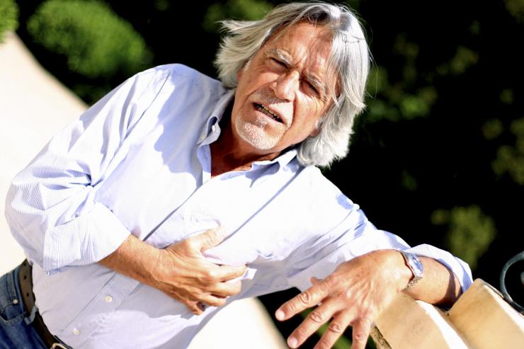
When something hurts, we try by all means to alleviate the condition and get rid of the pain. But it is not always possible to achieve the desired effect, and the reason for this is the lack of necessary knowledge. In order not to get lost in such situations, one must not only be able to identify possible cause disease, but also to know what measures to take.
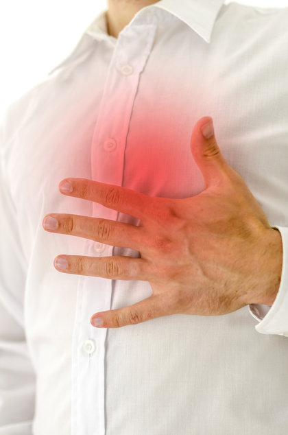
Most often, people are concerned about pain behind the sternum in the middle, which can be either the result of ordinary indigestion or a sign of the development of a dangerous disease. Having studied the symptoms of the most common diseases, you will know exactly what to do: undergo an examination at the clinic, solve the problem yourself, or call an ambulance at home.
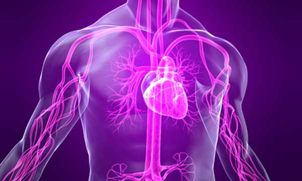
Most often, pain behind the sternum is associated with problems of the cardiovascular system. And in most cases, such assumptions are fully confirmed during the survey. Some forms of ischemic disease and aortic aneurysm are distinguished among the most serious pathologies.
Ischemia of the heart
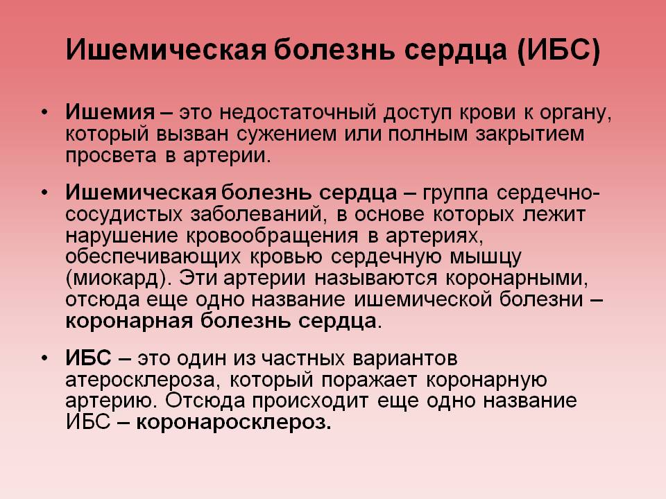
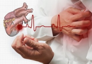
IHD (ischemic heart disease) is one of the most common causes of disability and death. Its development provokes a lack of oxygen in the heart muscle due to the narrowing of the coronary arteries. Despite all the advances in medicine, no means have yet been found to completely cure coronary artery disease. All known methods of treatment can only control the disease and slow down the development process. Depending on the degree of lack of oxygen and its duration, there are several forms of cardiac ischemia.
| Form of the disease | Characteristic manifestations |
|---|---|
| There are no obvious signs of the disease, narrowing of the arteries and the presence of atherosclerotic plaques can only be detected with an appropriate study |
| Chronic type of coronary artery disease, manifested by retrosternal pain with strong emotions and physical stress. Often accompanied by shortness of breath |
| Deterioration of the muscle. Each new attack is stronger than the previous one, additional symptoms may appear. As a rule, this form of the disease precedes a heart attack. |
| An acute condition often becomes chronic. The main manifestations are heart rhythm disturbances |
| An acute condition characterized by the death of a certain part of the heart muscle. Caused by complete blockage of an artery by a thrombus or plaque torn off the vessel wall |

Forms of coronary artery disease have different duration, intensity of development, often combined with each other. Depending on the individual characteristics of the organism, the course of the disease is acute or chronic.
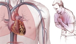
Symptoms of the disease:
- dull, pressing or sharp burning pain behind the sternum, radiating into the arm, under the shoulder blade, into the neck;
- shortness of breath during walking, climbing stairs, other physical exertion;
- frequent heartbeat, irregular heartbeat;
- increase in pressure;
- headache;
- the appearance of edema;
- pallor of the skin.
If you experience pain for the first time, you must immediately stop moving, sit down, and even better lie down and try to calm down, even out your breathing. If the room is cold, you need to cover yourself with a blanket, as hypothermia can also cause heart attacks. The pain usually goes away on its own within a minute.

For repeated attacks, it is advisable to have nitroglycerin on hand. As soon as the pain appears, you need to take a supine position, straighten up, put a pill under the tongue and hold until completely absorbed. If 5 minutes have passed and the pain has not disappeared, take another tablet. At one time, you can take no more than 5 tablets of nitroglycerin at five-minute intervals. If after that it does not get better, it is urgent to call an ambulance.
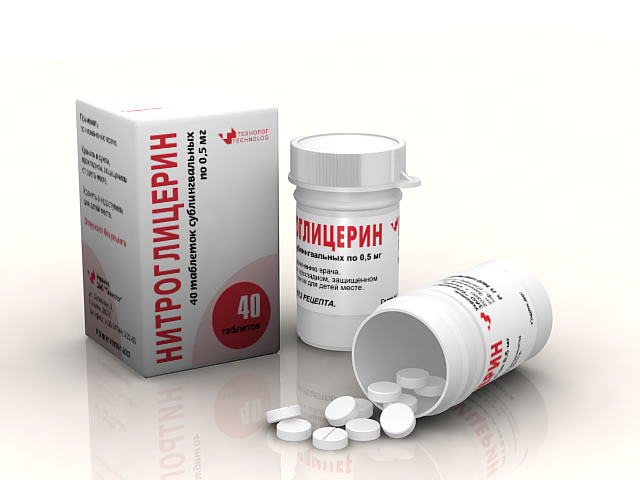
As a rule, pain manifestations of the chronic form of coronary artery disease are quickly removed with pills or drops. Aerosols act a little slower, but give a longer lasting effect.
![]()
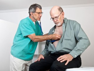
Here it is very important to notice in time the moment when the disease begins to progress: seizures become more frequent, shortness of breath appears faster when walking, to eliminate pain, not 1, but 2-3 tablets are required. Having found such signs, it is necessary to be examined by a cardiologist as soon as possible.
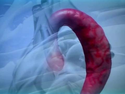
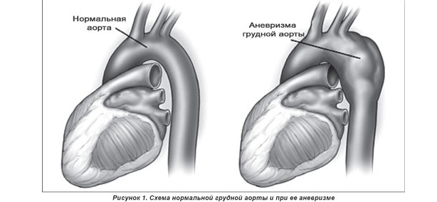
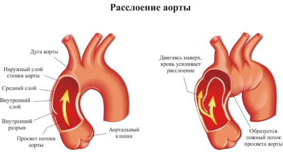
Aortic aneurysm is a dangerous disease. It is an expansion of individual sections of the aorta due to the thinning of the vascular walls. As a result, pressure on the walls of the aorta increases, fibrous tissues stretch, rupture and hemorrhage occur. As a rule, without the provision of qualified assistance, a person dies.
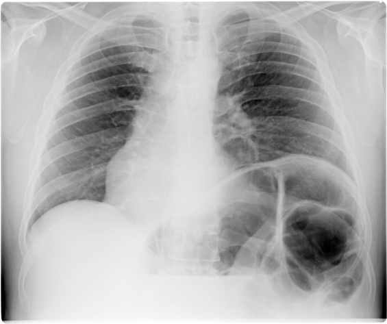
Aneurysms almost always develop asymptomatically, and this process can take years. Only at a late stage, when the blood vessel increases significantly and presses on adjacent organs, the patient begins to be disturbed by attacks of pain in different parts of the body. It is possible to detect an aneurysm using x-rays and ultrasound, examining the patient for other diseases. A timely detected pathology must be urgently treated, since a rupture can occur at any time.
Symptoms:
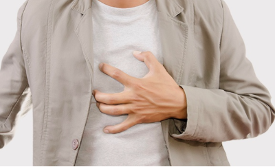
- very sharp, deep pain behind the sternum of a pulsating nature;
- back pain along the spine;
- shortness of breath and cough;
- pale skin;
- a sharp decrease in pressure;
- pulse asymmetry;
- darkening in the eyes;
- dizziness and weakness.
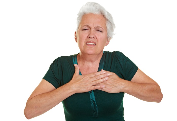
Sharp pain, pallor, and other symptoms of an aneurysm
What to do in such a situation? First of all, you need to call emergency help. Before the arrival of the doctor, the patient should lie down so that the upper part of the body is raised. It is impossible to move, as well as take any drugs - this can increase the hemorrhage. All further actions are taken by the doctor, the patient is hospitalized and the operation is performed.

With heart pain, you should reduce the load, avoid stressful situations as much as possible, give up coffee and bad habits. It is advisable to always have medicines with you, because it is not known when an attack will occur. If suddenly there was no nitroglycerin at hand, you can chew 1 aspirin tablet. You can’t get up, strain, walk until the pain disappears completely. And even after that, it’s better to lie down a little calmly for a while.
If there is no one around, and there are no medicines either, and the symptoms of an attack are already manifesting, use a very effective and simple method. You need to take a deep breath and cough hard, as if getting rid of sputum. Again a strong breath and cough, and so every 2 seconds for several minutes in a row.

What it does: when you inhale, the blood is saturated with oxygen, and coughing accelerates its circulation, causing heart contractions. Very often, this technique allows you to normalize the heart rhythm even before the ambulance arrives.
Autonomic disorders are most often observed in children and adolescents, and the causes of their occurrence include psycho-emotional factors, perinatal lesions nervous system, hereditary predisposition. Usually the disease is mild and treated on an outpatient basis. In rare cases, VVD acquires a severe degree, in which the patient's ability to work sharply decreases or is completely lost. Such patients are treated only permanently.
Symptoms:
- sudden attacks of retrosternal pain of a compressive or pressing character;
- cardiopalmus;
- suffocation;
- feeling of panic;
- pressure surges;
- low temperature;
- nausea and vomiting;
- stool disorders for no apparent reason;
- severe dizziness;
- sleep disorders;
- increasing lethargy;
- frequent depression.

Choking, panic, depression and other symptoms
In addition, many patients complain of constant cold feet and fingers, excessive sweating, and abdominal pain. During the examination, most of the physical parameters are within the normal range. Attacks can last from a few minutes to several days, and pain either increase or decrease. Usually the onset of an attack is preceded by a strong excitement or sudden physical exertion.

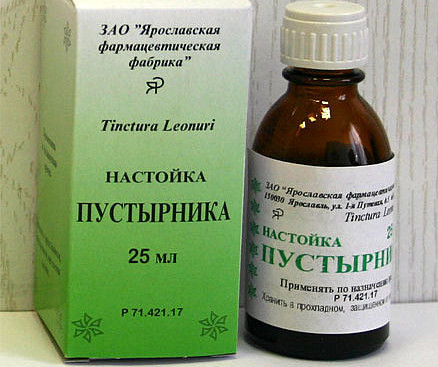
If you feel the approach of an attack, you need to take any sedative drug - validol, motherwort tincture, valerian, and find a quiet, calm place where you can lie down or at least sit comfortably.

Validol (Validol) - tablets

Try to breathe evenly and deeply, disconnect from all problems and external irritating factors. Self-massage of the head for several minutes helps to relieve tension. When the intensity of the attack begins to subside, you need to go to Fresh air and a little walk - this will improve well-being, reduce pain and tension. As soon as possible, it is necessary to be examined by a neurologist.
Pain in pathologies of the gastrointestinal tract
Pain in diseases of the stomach, intestines, certain types of hernias differ in nature from those of the heart, although they are localized in the chest area. Taking heart medications in this case has no effect, it can even aggravate the situation. To relieve a pain attack, you need to know what exactly causes it.
Diaphragmatic hernia
This type of hernia is characterized by the displacement of the peritoneal organs through the openings of the diaphragm into the chest cavity. Most often, this is part of the esophagus and the cardial part of the stomach, but intestinal loops can also be displaced. The cause of the pathology is congenital or acquired defects of the diaphragm, tissue weakness, regular overeating, hard work.
Symptoms:
- heartburn and frequent belching;
- moderate chest pain;
- fast saturation;
- vomit;
- rumbling and gurgling in the chest.
Heartburn, vomiting, pain behind the sternum - symptoms of diaphragmatic hernia
If the hernia is complicated by infringement, the person feels sudden pain in the left side of the sternum and abdomen, severe vomiting appears, and stool disorders may occur. This condition requires hospitalization and surgery. With a sliding hernia, surgery is not needed, the patient is simply prescribed a special diet with fractional nutrition, means to reduce acidity and reduce the production of gastric juice. In addition, it is necessary to exclude physical activity, wearing tight bandages or belts that squeeze the stomach and increase pressure inside the abdominal cavity.

To alleviate the condition, you should eat in small portions, sleep in a half-sitting position, putting 2 or 3 pillows under your head, and avoid sharp torso bends.

Take only those medicines prescribed by your doctor.

Gastritis and peptic ulcer disease are diagnosed in people of almost all age groups. With timely detection, these diseases can be successfully cured. One of common symptoms Both pathologies are pain in the chest, the attacks of which are sometimes very painful. Pain is accompanied by other symptoms:
- dyspepsia;
- belching;
- severe heartburn;
- a feeling of fullness and burning in the stomach;
- irritability;
- tachycardia.
![]()
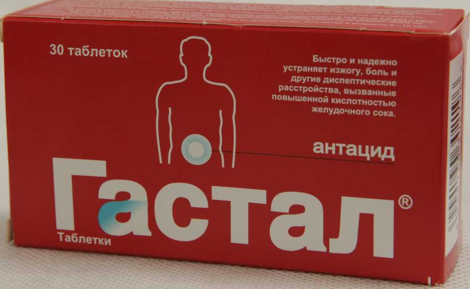
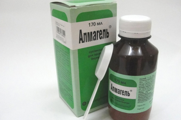
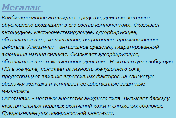
In case of an acute attack, it is best to call a doctor, in other cases, you can alleviate your well-being on your own. The most effective pain relievers are antacids, acid-neutralizing agents. These include Gastal, Rennie, Maalox, Almagel, Megalac and others.


 No-shpa
Help reduce pain and antispasmodics, for example, No-shpa and Papaverine (no more than 2 tablets). If there are no medicines at hand, and the pain is severe enough, you can use folk remedies. With an ulcer, a glass of warm milk or a small amount of semolina, a decoction of elecampane, an infusion of chamomile, celandine and yarrow quickly helps.


With gastritis, raw potato juice is very effective: 2 tubers are thoroughly washed, chopped in a meat grinder and the juice is squeezed out. Drink it on an empty stomach, an hour before meals, the course of treatment is 1 month.
![]()
Already after the first doses, the intensity of pain decreases. If the attack is strong, you need to clear the stomach: for this, they drink 2-3 glasses of warm water and induce vomiting. Further, rest, therapeutic fasting for 2 days, and plenty of fluids are recommended.
Osteochondrosis
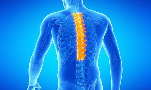
Chest pain is also among the symptoms. Damage to the discs between the vertebrae and pinching of the nerve roots in the thoracic region of the spine cause pain similar to heart pain. This often leads to misdiagnosis and complicates treatment.
It is possible to distinguish osteochondrosis from other diseases by some specific features, among which:
- numbness and strain of the back muscles;
- increased pain when bending and turning the torso, raising arms, hypothermia, and also at night;
- pain occurs when you take a deep breath;
- a feeling of tingling and "goosebumps" in certain parts of the body;
- tightness in the chest;
- pain between the ribs when walking;
- cold or burning sensation in the legs.

Taking nitroglycerin or other cardiac drugs does not give any effect, which is also evidence of the neuralgic origin of pain. Changing the position of the body helps to alleviate the condition - the pain does not disappear completely, but its intensity decreases. For treatment, medicines, acupuncture, traction, therapeutic massage and special gymnastics are used.
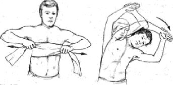
To cure osteochondrosis, you need to be patient. Proper nutrition means a lot, with an emphasis on foods containing magnesium, calcium and vitamins.
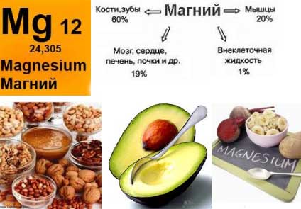
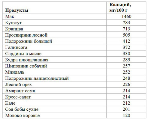
These are seafood, nuts, legumes, spinach and cabbage, fresh milk, bran bread. Such nutrition at the same time will help to lose excess weight, which puts additional stress on the vertebrae. Regular moderate exercise is no less important: daily exercises strengthen the back and reduce the risk of complications for the spine.
Video - Pain behind the sternum in the middle: what to do
When pain in the middle of the chest is disturbing, this may indicate the presence of both a simple overstrain of the body, or fatigue, and the presence of serious diseases. In any case, it is necessary to find out the cause of their appearance and eliminate it by visiting a doctor in a timely manner.
Causes and manifestations of chest pain
The sensations of pain that occur in the middle of the chest are familiar to the vast majority of mankind. The reasons for this can be very diverse. It is good when it is the cause of simple physical overexertion, but worse if chest pain indicates the presence of a serious illness.
Chest pain can be caused by a disease of the organs directly in the chest, or indicate diseases of the organs located in the abdominal cavity. In addition, diseases of the diaphragm, esophagus, lungs or heart can also cause pain in the middle of the chest.
But no matter what it is called, one way or another, chest pain in the middle always gives a reason to worry about your health. First of all, it is necessary to find out the causes of such pain, and to exclude the diseases by which it can be caused. Such work is only possible for qualified specialists in the field of medicine.
Turning to the doctor, you need to know the nature of the pain that accompanies you in the middle of the chest. Depending on it, the pain manifests itself in the following sensations:
aching pain in the middle of the chest;
dull chest pain;
sharp pain;
burning pain in the chest;
pressing pain in middle of chest.
If you experience any of the above pain in the chest, you should immediately consult a doctor.
Chest pain can be caused by both minor and dangerous causes.
Dangerous diseases that can cause pain in the middle of the chest
Because chest pain is different character, it can be caused by one of the following dangerous and serious diseases.
aortic dissection;
ischemic disease of the organ - the heart;
any kind of heart attack;
thromboembology of the lung system;
oncological diseases of the respiratory organs, heart, spine;
rupture of an ulcer of the duodenum or stomach;
acute pancreatitis
At the first appearance of discomfort in the middle of the chest, you should not delay visiting a doctor. A quick visit will allow you to identify serious diseases in the early stages and eliminate them. If the chest pain is pressing or burning, it is necessary to call a doctor, as these may be manifestations of an angina attack.

Chest pain is a reason to visit a doctor
With a well-defined duration of chest pain, a heart attack often manifests itself. You should not refuse hospitalization even if, upon arrival of the doctors, the pain disappeared, and the electrocardiogram showed a negative result.
Often, diseases that are psychogenic in nature cause pain in the middle of the chest. Symptoms of such diseases can be sharp, stabbing, pressing and dull pain. Usually, pain in such diseases manifests itself in the left upper chest, but sometimes it is also felt in the middle of the chest.
You can distinguish a psychological illness that provoked chest pain by the following symptoms:
The nature of the pain, its localization and intensity often change.
The pain is quite long, after taking sedatives it subsides.
If chest pain is persistent
If chest pain is felt constantly, this may indicate that it is caused by less dangerous diseases, compared with pain that appears suddenly. Such pain can indicate injuries and diseases of the spine or neurological diseases.
In addition, the feeling of constant pain in the middle of the chest that does not subside can be caused by diseases such as:
disorders of the pancreas;
stomach diseases;
diseases of the digestive tract.
If constant pain in the middle of the chest increases with time, this indicates that the disease that caused them is progressing and developing.
Pain in the middle of the chest, even the most insignificant, should not go unnoticed. When they appear, a visit to the doctor is essential. If the disease, even if not too serious, is just beginning, a timely visit to a specialist will allow you to get rid of it at an early stage, and will not allow the disease to develop into more serious and dangerous forms.
The main causes of pain between the breasts:
- diseases of the musculoskeletal system: costal chondritis, rib fracture;
- cardiovascular diseases: cardiac ischemia caused by atherosclerosis of the heart vessels; unstable / stable angina; cardiac ischemia caused by coronary vasospasm (angina pectoris); mitral valve prolapse syndrome; cardiac arrhythmia; pericarditis.
- gastrointestinal diseases: gastroesophageal reflux, spasm of the esophagus, gastric and duodenal ulcer, gallbladder disease;
- anxiety states: vague anxiety or "stress", panic disorder;
- pulmonary diseases: pleurodynia (pleuralgia), acute bronchitis, pneumonia;
- neurological diseases;
- uncharacteristic defined or atypical pain between the breasts.
Pain between the breasts is not limited to a certain age group, but is more common in adults than in children. The highest percentage is observed among adults over 65 years of age, and in second place are male patients aged 45 to 65 years.
Frequency of diagnosis, by age and sex
|
Age group (years) |
The most common diagnoses |
|
|
1. Gastroesophageal reflux |
||
|
2. Muscular pain of the chest wall |
||
|
3. Costal chondritis |
||
|
2. Muscular pain of the chest wall |
||
|
65 and more |
||
|
2. "Atypical" pain between the breasts or coronary artery disease |
||
|
1. Costal chondritis |
||
|
2. Anxiety/stress |
||
|
1. Muscular pain of the chest wall |
||
|
2. Costal chondritis |
||
|
3. "Atypical" pain between the breasts |
||
|
4. Gastroesophageal reflux |
||
|
1. Angina, unstable angina, myocardial infarction |
||
|
2. "Atypical" pain between the breasts |
||
|
3. Muscular pain of the chest wall |
||
|
65 and more |
1. Angina, unstable angina, myocardial infarction |
|
|
2. Muscular pain of the chest wall |
||
|
3. "Atypical" pain between the breasts or costal chondritis |
No less difficult is the position of the doctor in the initial interpretation of pain, when he tries to connect it with the pathology of one or another organ. Observation of clinicians of the last century helped them formulate assumptions about the pathogenesis of pain - if an attack of pain occurs without a reason and stops on its own, then the pain is likely to be functional. Works dedicated to detailed analysis pain between the breasts, not numerous; the groupings of pains proposed in them are far from perfect. These shortcomings are due to objective difficulties in analyzing the patient's sensations.
The complexity of interpreting pain in the chest is also due to the fact that the detected pathology of one or another organ of the chest or musculoskeletal formation does not mean that it is the source of pain; in other words, the identification of the disease does not mean that the cause of pain is accurately determined.
When evaluating patients with pain between the breasts, the clinician must weigh all relevant options for potential causes of pain, determine when intervention is needed, and choose from a virtually limitless number of diagnostic and therapeutic strategies. All this must be done while responding to the distress experienced by patients concerned about the presence of a life-threatening illness. The difficulty in diagnosis is further complicated by the fact that pain between the breasts is often a complex interplay of psychological, pathological, and psychosocial factors. This makes it the most common problem in primary care.
When considering pain between the breasts, the following five elements should be considered (at a minimum): predisposing factors; description of the attack of pain; duration of painful episodes; characterization of the actual pain; pain relieving factors.
With all the variety of causes that cause pain in the chest, pain syndromes can be grouped.
Approaches to groupings may be different, but basically they are built according to the nosological or organ principle.
Conventionally, 6 following groups of causes of pain between the breasts can be distinguished:
- Pain caused by heart disease (so-called heart pain). These pains can be the result of damage or dysfunction of the coronary arteries - coronary pain. The "coronary component" does not take part in the origin of non-coronary pain. In the future, we will use the terms "heart pain syndrome", "heart pain", understanding their connection with a particular pathology of the heart.
- Pain caused by pathology of large vessels (aorta, pulmonary artery and its branches).
- Pain caused by pathology of the bronchopulmonary apparatus and pleura.
- Pain associated with the pathology of the spine, anterior chest wall and muscles of the shoulder girdle.
- Pain caused by pathology of the mediastinal organs.
- Pain associated with diseases of the abdominal organs and pathology of the diaphragm.
Pain in the chest area is also divided into acute and long-term, with an obvious cause and without an apparent cause, "non-dangerous" and pain that is a manifestation of life-threatening conditions. Naturally, first of all it is necessary to establish whether the pain is dangerous or not. "Dangerous" pains include all types of anginal (coronary) pains, pain in pulmonary embolism (PE), dissecting aortic aneurysm, spontaneous pneumothorax. By "non-dangerous" - pain in the pathology of the intercostal muscles, nerves, bone and cartilage formations of the chest. "Dangerous" pains are accompanied by a suddenly developed serious condition or severe disorders of the heart or respiratory function, which immediately allows you to narrow the range of possible diseases (acute myocardial infarction, pulmonary embolism, dissecting aortic aneurysm, spontaneous pneumothorax).
The main causes of acute pain between the breasts, which are life-threatening:
- cardiological: acute or unstable angina pectoris, myocardial infarction, dissecting aortic aneurysm;
- pulmonary: pulmonary embolism; tension pneumothorax.
It should be noted that the correct interpretation of pain between the breasts is quite possible with the usual physical examination of the patient using a minimum number of instrumental methods (conventional electrocardiographic and x-ray examination). An erroneous initial idea of the source of pain, in addition to increasing the period of examination of the patient, often leads to serious consequences.
History and physical examination findings to determine the causes of pain between the breasts
|
History data |
|||
|
Cardiac |
Gastrointestinal |
Musculoskeletal |
|
|
Predisposing factors |
Male gender. Smoking. Increased blood pressure. Hyperlipidemia. Family history of myocardial infarction |
Smoking. Alcohol consumption |
Physical activity. New type of activity. Abuse. Recurring actions |
|
Characteristics of an attack of pain |
With high levels of stress or emotional stress |
After eating and/or on an empty stomach |
During activity or after |
|
Pain duration |
Several minutes to several hours |
From hours to days |
|
|
Characteristics of pain |
Pressure or "burning" |
Pressure or boring pain |
Acute, localized, caused by movements |
|
Pain relief factors |
Nitropreparations under the tongue |
Taking food. Antacids. Antihistamines |
Rest. Analgesics. Non-steroidal anti-inflammatory drugs |
|
Supporting data |
With angina attacks, rhythm disturbances or noises are possible |
Soreness in the epigastric region |
Pain on palpation in the paravertebral points, at the exit of the intercostal nerves, soreness of the periosteum |
Cardialgia (non-anginal pain). Cardialgia, caused by certain diseases of the heart, are very common. By its origin, significance and place in the structure of the incidence of the population, this group of pains is extremely heterogeneous. The causes of such pain and their pathogenesis are very diverse. Diseases or conditions in which cardialgia is observed are as follows:
- Primary or secondary cardiovascular functional disorders - the so-called cardiovascular syndrome of the neurotic type or neurocirculatory dystonia.
- Diseases of the pericardium.
- Inflammatory diseases of the myocardium.
- Dystrophy of the heart muscle (anemia, progressive muscular dystrophy, alcoholism, beriberi or starvation, hyperthyroidism, hypothyroidism, catecholamine effects).
As a rule, non-anginal pains are benign, as they are not accompanied by coronary insufficiency and do not lead to the development of ischemia or myocardial necrosis. However, in patients with functional disorders leading to an increase (usually short-term) in the level of biologically active substances (catecholamins), the likelihood of ischemia still exists.
Pain between the breasts of neurotic origin. We are talking about pain in the region of the heart, as one of the manifestations of neurosis or neurocirculatory dystonia (vegetative-vascular dystonia). Usually these are pains of a aching or stabbing nature, of varying intensity, sometimes long-term (hours, days) or, conversely, very short-term, instantaneous, penetrating. The localization of these pains is very different, not always constant, almost never retrosternal. Pain may increase with physical exertion, but usually with psycho-emotional stress, fatigue, without a clear effect of the use of nitroglycerin, they do not decrease at rest, and, sometimes, on the contrary, patients feel better when moving. The diagnosis takes into account the presence of signs of a neurotic state, autonomic dysfunction (sweating, dermographism, subfebrile condition, pulse and blood pressure fluctuations), as well as young or average age patients, mostly females. These patients have increased fatigue, decreased exercise tolerance, anxiety, depression, phobias, fluctuations in pulse, blood pressure. In contrast to the severity of subjective disorders, an objective study, including using various additional methods, does not reveal a specific pathology.
Sometimes among these symptoms of neurotic origin, the so-called hyperventilation syndrome is revealed. This syndrome is manifested by an arbitrary or involuntary increase and deepening of respiratory movements, tachycardia, arising in connection with adverse psycho-emotional influences. This may cause pain between the breasts, as well as paresthesia and muscle twitching in the limbs due to the resulting respiratory alkalosis. There are observations (incompletely confirmed) indicating that hyperventilation can lead to a decrease in myocardial oxygen consumption and provoke coronary spasm with pain and ECG changes. It is possible that it is hyperventilation that can cause pain in the region of the heart during exercise testing in individuals with vegetative-vascular dystonia.
To diagnose this syndrome, a provocative test with induced hyperventilation is performed. The patient is asked to breathe more deeply - 30-40 times per minute for 3-5 minutes or until the patient's usual symptoms appear (pain between the breasts, headaches, dizziness, shortness of breath, sometimes fainting). The appearance of these symptoms during the test or 3-8 minutes after its completion, with the exclusion of other causes of pain, has a definite diagnostic value.
Hyperventilation in some patients may be accompanied by aerophagia with the appearance of pain or a feeling of heaviness in the upper part of the epigastric region due to distension of the stomach. These pains can spread upward, behind the sternum, into the neck and the area of the left shoulder blade, simulating angina pectoris. Such pains are aggravated by pressure on the epigastric region, in the supine position, with deep breathing, they decrease with belching with air. With percussion, an expansion of the Traube space zone is found, including tympanitis over the area of \u200b\u200babsolute dullness of the heart, with fluoroscopy - an enlarged gastric bladder. Similar pains can occur when stretching the gases of the left corner of the colon. In this case, the pain is often associated with constipation and is relieved after a bowel movement. A thorough history usually allows the true nature of the pain to be determined.
The pathogenesis of cardiac pain in neurocirculatory dystonia is unclear, due to the impossibility of their experimental reproduction and confirmation in the clinic and experiment, in contrast to anginal pain. Perhaps, in connection with this circumstance, a number of researchers generally question the presence of pain in the heart with neurocirculatory dystonia. Similar trends are most common among representatives of the psychosomatic trend in medicine. According to their views, we are talking about the transformation of psycho-emotional disorders into pain.
The origin of pain in the heart in neurotic states is also explained from the standpoint of the cortico-visceral theory, according to which, when the vegetative devices of the heart are stimulated, a pathological dominant appears in the central nervous system with the formation of a vicious circle. There is reason to believe that pain in the heart with neurocirculatory dystonia occurs due to a violation of myocardial metabolism against the background of excessive adrenal stimulation. At the same time, a decrease in the content of intracellular potassium, activation of dehydrogenation processes, an increase in the level of lactic acid and an increase in myocardial oxygen demand are observed. Hyperlactatemia is a well-proven fact in neurocirculatory dystonia.
Clinical observations indicating a close relationship between pain in the region of the heart and emotional influences confirm the role of catecholamines as a trigger for pain. This position is supported by the fact that intravenous administration of izadrin to patients with neurocirculatory dystonia causes pain in the region of the heart such as cardialgia. Obviously, catecholamine stimulation can also explain the provocation of cardialgia with a test with hyperventilation, as well as its occurrence at the height of respiratory disorders with neurocirculatory dystonia. This mechanism can also be confirmed positive results treatment of cardialgia with breathing exercises aimed at eliminating hyperventilation. A certain role in the formation and maintenance of pain in the heart syndrome in neurocirculatory dystonia is played by the flow of pathological impulses coming from hyperalgesia zones in the muscles of the anterior chest wall to the corresponding segments of the spinal cord, where, according to the "gateway" theory, the summation phenomenon occurs. In this case, a reverse flow of impulses is noted, causing irritation of the thoracic sympathetic ganglia. Of course, the low threshold of pain sensitivity in vegetative-vascular dystonia also matters.
In the occurrence of pain, such still insufficiently studied factors as impaired microcirculation, changes in the rheological properties of the blood, and an increase in the activity of the kinincallikrein system can play a role. It is possible that with the long-term existence of severe vegetative-vascular dystonia, its transition to coronary artery disease with unchanged coronary arteries, in which pain is caused by spasm of the coronary arteries, is possible. In a targeted study of a group of patients with proven coronary artery disease with unchanged coronary arteries, it was found that all of them suffered from severe neurocirculatory dystonia in the past.
In addition to vegetative-vascular dystonia, cardialgia is also observed in other diseases, but the pain is less pronounced and usually never comes to the fore in the clinical picture of the disease.
The origin of pain in pericardial lesions is quite understandable, since there are sensitive nerve endings in the pericardium. Moreover, it has been shown that irritation of certain areas of the pericardium gives different localization of pain. For example, irritation of the pericardium on the right causes pain along the right mid-clavicular line, and irritation of the pericardium in the region of the left ventricle is accompanied by pain spreading along the inner surface of the left shoulder.
Pain in myocarditis of various origins is a very common symptom. Their intensity is usually low, but in 20% of cases they have to be differentiated from pain caused by coronary artery disease. Pain in myocarditis is probably associated with irritation of the nerve endings located in the epicardium, as well as with inflammatory myocardial edema (in the acute phase of the disease).
Even more uncertain is the origin of pain in myocardial dystrophies of various origins. Probably, the pain syndrome is due to a violation of myocardial metabolism, the concept of local tissue hormones, convincingly presented by N.R. Paleev et al. (1982) may also shed light on the causes of pain. In some myocardial dystrophies (due to anemia or chronic carbon monoxide poisoning), pain can be of mixed origin, in particular, the ischemic (coronary) component is essential.
It is necessary to dwell on the analysis of the causes of pain in patients with myocardial hypertrophy (due to pulmonary or systemic hypertension, valvular heart disease), as well as in primary cardiomyopathies (hypertrophic and dilated). Formally, these diseases are mentioned in the second heading of anginal pain caused by an increase in myocardial oxygen demand with unchanged coronary arteries (the so-called non-coronary forms). However, under these pathological conditions, in some cases, unfavorable hemodynamic factors occur, causing relative myocardial ischemia. It is believed that the angina-type pain observed in aortic insufficiency depends primarily on low diastolic pressure and, consequently, low coronary perfusion (coronary blood flow is realized during diastole).
With aortic stenosis or idiopathic myocardial hypertrophy, the appearance of pain is associated with impaired coronary circulation in the subendocardial regions due to a significant increase in intramyocardial pressure. All pain sensations in these diseases can be designated as metabolically or hemodynamically caused anginal pain. Despite the fact that they do not formally refer to IHD, one should keep in mind the possibility of developing small-focal necrosis. However, the characteristics of these pains often do not correspond to classical angina pectoris, although typical attacks are also possible. In the latter case, the differential diagnosis with CAD is particularly difficult.
In all cases of detection of non-coronary causes of the origin of pain between the breasts, it is taken into account that their presence does not at all contradict the simultaneous existence of IHD and, accordingly, requires an examination of the patient in order to exclude or confirm it.
Pain between the breasts due to pathology of the bronchopulmonary apparatus and pleura. Pain often accompanies a variety of pulmonary pathologies, occurring both in acute and in chronic diseases. However, it is usually not a leading clinical syndrome and is quite easily differentiated.
The source of pain is the parietal pleura. From pain receptors located in the parietal pleura, afferent fibers go as part of the intercostal nerves, so the pain is clearly localized on the affected half of the chest. Another source of pain is the mucous membrane of the large bronchi (which is well proven with bronchoscopy) - afferent fibers from the large bronchi and trachea are part of the vagus nerve. The mucous membrane of the small bronchi and lung parenchyma probably does not contain pain receptors, so pain in the primary lesion of these formations appears only when the pathological process (pneumonia or tumor) reaches the parietal pleura or spreads to large bronchi. The most severe pains are noted during the destruction of the lung tissue, sometimes acquiring high intensity.
The nature of pain sensations to some extent depends on their origin. Pain in the parietal pleura is usually stabbing, clearly associated with coughing and deep breathing. Dull pain is associated with distension of the mediastinal pleura. Severe constant pain, aggravated by breathing, moving the arms and shoulder girdle, may indicate tumor growth into the chest.
Most common causes pulmonary-pleural pains are pneumonia, lung abscess, tumors of the bronchi and pleura, pleurisy. With pain associated with pneumonia, dry or exudative pleurisy, auscultation may reveal wheezing in the lungs, pleural friction noise.
Severe pneumonia in adults has the following clinical features:
- moderate or severe respiratory depression;
- temperature 39.5 °C or higher;
- confusion;
- respiratory rate - 30 per minute or more;
- pulse 120 beats per minute or more;
- systolic blood pressure below 90 mm Hg. Art.;
- diastolic blood pressure below 60 mm Hg. Art.;
- cyanosis;
- over 60 years old - features: confluent pneumonia, is more severe with concomitant severe diseases (diabetes, heart failure, epilepsy).
NB! All patients with signs of severe pneumonia should be immediately referred to hospital! Referral to hospital:
- severe form of pneumonia;
- patients with pneumonia from socioeconomically disadvantaged backgrounds, or who are unlikely to follow doctor's orders at home; who live very far from a medical facility;
- pneumonia in combination with other diseases;
- suspicion of atypical pneumonia;
- patients who do not respond well to treatment.
Pneumonia in children is described as follows:
- retraction of the intercostal spaces of the chest, cyanosis and inability to drink in young children (from 2 months to 5 years) is also a sign of severe pneumonia, which requires an urgent referral to a hospital;
- pneumonia should be distinguished from bronchitis: the most valuable symptom in the case of pneumonia is tachypnea.
Pain in case of damage to the pleura almost does not differ from those in acute intercostal myositis or trauma to the intercostal muscles. With spontaneous pneumothorax, there is an acute unbearable pain between the breasts, associated with damage to the bronchopulmonary apparatus.
Pain between the breasts, difficult to interpret due to its vagueness and isolation, is observed in initial stages bronchogenic lung cancer. The most excruciating pain is characteristic of the apical localization of lung cancer, when damage to the common trunk of CVII and ThI nerves and the brachial plexus almost inevitably and rapidly develops. The pain is localized mainly in the brachial plexus and radiates along the outer surface of the arm. On the side of the lesion, Horner's syndrome (narrowing of the pupil, ptosis, enophthalmos) often develops.
Pain syndromes also occur with mediastinal localization of cancer, when compression of the nerve trunks and plexuses causes acute neuralgic pain in the shoulder girdle, upper limb, and chest. This pain gives rise to an erroneous diagnosis of angina pectoris, myocardial infarction, neuralgia, plexitis.
The need for differential diagnosis of pain caused by damage to the pleura and bronchopulmonary apparatus, with coronary artery disease arises in cases where the picture of the underlying disease is fuzzy and pain comes to the fore. In addition, such differentiation (especially in acute unbearable pain) should be carried out with diseases caused by pathological processes in large vessels - pulmonary embolism, dissecting aneurysm of various parts of the aorta. Difficulties in identifying pneumothorax as a cause of acute pain are due to the fact that in many cases the clinical picture of this acute situation is erased.
Pain between the breasts associated with the pathology of the mediastinal organs is caused by diseases of the esophagus (spasm, reflux esophagitis, diverticula), mediastinal tumors and mediastinitis.
Pain in diseases of the esophagus usually have a burning character, are localized in the chest, occur after eating, and are aggravated in a horizontal position. Such usual symptoms as heartburn, belching, and swallowing disorders may be absent or be mildly pronounced, and retrosternal pain, which often occurs during exercise and is inferior to the action of nitroglycerin, comes to the fore. The similarity of these pains with angina pectoris is complemented by the fact that they can radiate to the left half of the chest, shoulders, arms. With a more detailed questioning, however, it turns out that the pains are more often associated with food, especially plentiful, and not with physical activity, usually occur in the supine position and disappear or are relieved when moving to a sitting or standing position, when walking, after taking antacids, for example, soda, which is uncharacteristic for coronary artery disease. Often, palpation of the epigastric region increases these pains.
Retrosternal pain is also suspicious for gastroesophageal reflux and esophagitis. to confirm the presence of which 3 types of tests are important: endoscopy and biopsy; intraesophageal infusion of 0.1% hydrochloric acid solution; monitoring intraesophageal pH. Endoscopy is important to detect reflux, esophagitis, and to rule out other pathologies. X-ray examination of the esophagus with barium reveals anatomical changes, but its diagnostic value is considered relatively low due to the high frequency of false positive signs of reflux. With the perfusion of hydrochloric acid (120 drops per minute through a probe), the appearance of the usual pains for the patient matters. The test is considered to be highly sensitive (80%), but not specific enough, which requires repeated studies in case of fuzzy results.
If the results of endoscopy and perfusion of hydrochloric acid are unclear, monitoring of intraesophageal pH can be carried out using a radio telemetry capsule placed in the lower part of the esophagus for 24-72 hours. indeed a criterion for the esophageal origin of pain.
Pain between the breasts, similar to angina pectoris, can also be a consequence of an increase in the motor function of the esophagus with achalasia (spasm) of the cardiac section or diffuse spasm. Clinically, in such cases, there are usually signs of dysphagia (especially when taking solid food, cold liquids), which, unlike organic stenosis, is unstable. Sometimes retrosternal pains of different duration come to the fore. Difficulties in differential diagnosis are also due to the fact that this category of patients is sometimes helped by nitroglycerin, which relieves spasm and pain.
Radiographically, with achalasia of the esophagus, an expansion of its lower part and a delay in it of the barium mass are detected. However, an x-ray examination of the esophagus in the presence of pain is of little information, or rather, of little evidence: false positive results were noted in 75% of cases. Esophageal manometry using a three-lumen probe is more effective. The coincidence in time of onset of pain and increased intraesophageal pressure has a high diagnostic value. In such cases, there may be a positive effect of nitroglycerin and calcium antagonists, which reduce the tone of smooth muscles and intraesophageal pressure. Therefore, these drugs can be used in the treatment of such patients, especially in combination with anticholinergics.
Clinical experience suggests that coronary artery disease is indeed often misdiagnosed in esophageal pathology. In order to make a correct diagnosis, the doctor must look for other symptoms of esophageal disorders in the patient and compare the clinical manifestations and the results of various diagnostic tests.
Attempts to develop a set of instrumental studies that would help distinguish between anginal and esophageal pain were unsuccessful, since this pathology is often combined with angina pectoris, which is confirmed by bicycle ergometry. Thus, despite the use of various instrumental methods, the differentiation of pain sensations is still very difficult.
Mediastinitis and tumors of the mediastinum are infrequent causes of pain between the breasts. Usually, the need for differential diagnosis with coronary artery disease arises at pronounced stages of tumor development, when, however, there are still no pronounced symptoms of compression. The appearance of other signs of the disease greatly facilitates the diagnosis.
Pain between the breasts in diseases of the spine. Pain in the chest can also be associated with degenerative changes in the spine. The most common disease of the spine is osteochondrosis (spondylosis) of the cervical and thoracic region, in which there is pain, sometimes similar to angina pectoris. This pathology is widespread, since changes in the spine are often observed after 40 years. With damage to the cervical and (or) upper thoracic spine, the development of a secondary radicular syndrome with the spread of pain in the chest area is often observed. These pains are associated with irritation of the sensory nerves by osteophytes and thickened intervertebral discs. Usually, bilateral pains appear in the corresponding intercostal spaces, but patients quite often concentrate their attention on their retrosternal or pericardial localization, referring them to the heart. Such pains can be similar to angina pectoris in the following ways: they are perceived as a feeling of pressure, heaviness, sometimes radiate to the left shoulder and arm, neck, can be provoked by physical activity, accompanied by a feeling of shortness of breath due to the impossibility of deep breathing. Taking into account the advanced age of patients in such cases, the diagnosis of coronary artery disease is often made with all the ensuing consequences.
At the same time, degenerative changes in the spine and the pain caused by them can also be observed in patients with undoubted coronary artery disease, which also requires a clear distinction between the pain syndrome. Perhaps, in some cases, angina pectoris against the background of atherosclerosis of the coronary arteries in patients with spinal lesions occur reflexively. The unconditional recognition of such a possibility, in turn, shifts the "center of gravity" to the pathology of the spine, reducing the importance of independent damage to the coronary arteries.
How to avoid diagnostic errors and make the correct diagnosis? Of course, it is important to conduct an X-ray of the spine, but the changes detected in this case are completely insufficient for diagnosis, since these changes can only accompany IHD and (or) not be clinically manifested. Therefore, it is very important to find out all the features of pain. As a rule, pain depends not so much on physical activity as on changes in body position. Pain is often aggravated by coughing, deep breathing, may decrease in some comfortable position of the patient, after taking analgesics. These pains differ from angina pectoris in a more gradual onset, longer duration, they do not go away at rest and after the use of nitroglycerin. Irradiation of pain in left hand occurs on the dorsal surface, in the I and II fingers, while with angina pectoris - in the IV and V fingers of the left hand. Of certain importance is the detection of local pain in the spinous processes of the corresponding vertebrae (trigger zone) when pressed or tapped paravertebral and along the intercostal space. Pain can also be caused by certain techniques: strong pressure on the head towards the back of the head or stretching one arm while turning the head to the other side. With bicycle ergometry, pain in the region of the heart may appear, but without characteristic ECG changes.
Thus, the diagnosis of radicular pain requires a combination of radiological signs of osteochondrosis and characteristic features of pain between the breasts that do not correspond to coronary artery disease.
The frequency of muscular-fascial (muscular-dystonic, muscular-dystrophic) syndromes in adults is 7-35%, and in some professional groups it reaches 40-90%. In some of them, heart disease is often misdiagnosed, since the pain syndrome in this pathology has some similarities with pain in cardiac pathology.
There are two stages of the disease of muscular-fascial syndromes (Zaslavsky E.S., 1976): functional (reversible) and organic (muscular-dystrophic). In the development of muscular-fascial syndromes, there are several etiopathogenetic factors:
- Injuries of soft tissues with the formation of hemorrhages and sero-fibrinous extravasates. As a result, compaction and shortening of the muscles or individual muscle bundles, ligaments, and a decrease in the elasticity of the fascia develop. As a manifestation of an aseptic inflammatory process, connective tissue is often formed in excess.
- Microtraumatization of soft tissues in some types of professional activity. Microtraumas disrupt tissue circulation, cause muscular-tonic dysfunction with subsequent morphological and functional changes. This etiological factor is usually combined with others.
- Pathological impulsation in visceral lesions. This impulse, which occurs when the internal organs are damaged, is the cause of the formation of various sensory, motor and trophic phenomena in the integumentary tissues, innervationally associated with the altered internal organ. Pathological interoceptive impulses, switching through the spinal segments, go to the corresponding affected internal organ - connective tissue and muscle segments. The development of muscular-fascial syndromes associated with cardiovascular pathology can change the pain syndrome so much that diagnostic difficulties arise.
- Vertebrogenic factors. When receptors of the affected motor segment are irritated (receptors of the fibrous ring of the intervertebral disc, posterior longitudinal ligament, joint capsules, autochthonous muscles of the spine), not only local pain and muscle-tonic disorders occur, but also various reflex responses at a distance - in the area of integumentary tissues, innervationally connected with affected vertebral segments. But far from all cases, there is a parallelism between the severity of radiographic changes in the spine and clinical symptoms. Therefore, the radiographic signs of osteochondrosis cannot yet serve as an explanation for the development of muscular-fascial syndromes solely by vertebrogenic factors.
As a result of the influence of several etiological factors, muscular-tonic reactions develop in the form of hypertonicity of the affected muscle or muscle group, which is confirmed by electromyography. Muscle spasm is one of the sources of pain. In addition, impaired microcirculation in the muscle leads to local tissue ischemia, tissue edema, accumulation of kinins, histamine, and heparin. All these factors also cause pain. If muscular-fascial syndromes are observed for a long time, then fibrous degeneration of muscle tissue occurs.
The greatest difficulties in the differential diagnosis of muscular-fascial syndromes and pain of cardiac origin are found in the following syndromes: shoulder-scapular periarthritis, scapular-costal syndrome, anterior chest wall syndrome, interscapular pain syndrome, pectoralis minor syndrome, anterior scalene syndrome. Anterior chest wall syndrome is observed in patients after myocardial infarction, as well as in non-coronary heart lesions. It is assumed that after a myocardial infarction, the flow of pathological impulses from the heart spreads through the segments of the vegetative chain and leads to degenerative changes in the corresponding formations. This syndrome in persons with a known healthy heart may be due to traumatic myositis.
More rare syndromes, accompanied by pain in the anterior chest wall, are: Tietze's syndrome, xifoidia, manubriosternal syndrome, scalenus syndrome.
Tietze's syndrome is characterized by severe pain at the junction of the sternum with the cartilages of the II-IV ribs, swelling of the costal-cartilaginous joints. It is observed mainly in middle-aged people. Etiology and pathogenesis are unclear. There is an assumption about aseptic inflammation of the costal cartilages.
Xifoidia is manifested by a sharp pain between the breasts, aggravated by pressure on the xiphoid process, sometimes accompanied by nausea. The cause of the pain is unclear, perhaps there is a connection with the pathology of the gallbladder, duodenum, stomach.
With manubriosternal syndrome, acute pain is noted above the upper part of the sternum or somewhat laterally. The syndrome is observed in rheumatoid arthritis, but occurs in isolation, and then it becomes necessary to differentiate it from angina pectoris.
Scalenus syndrome - compression of the neurovascular bundle of the upper limb between the anterior and middle scalene muscle, as well as the normal I or additional rib. Pain in the region of the anterior chest wall is combined with pain in the neck, shoulder girdle, shoulder joints, sometimes there is a wide area of irradiation. At the same time, vegetative disorders are observed in the form of chills, pallor of the skin. Difficulty breathing, Raynaud's syndrome.
Summarizing the above, it should be noted that the true frequency of pain of this origin is unknown, therefore, it is not possible to determine their specific weight in the differential diagnosis of angina pectoris.
Differentiation is necessary in the initial period of the disease (when they first think about angina) or if the pain caused by the listed syndromes is not combined with other signs that allow them to correctly recognize their origin. At the same time, pains of this origin can be combined with true coronary artery disease, and then the doctor must also understand the structure of this complex pain syndrome. The need for this is obvious, since the correct interpretation will affect both treatment and prognosis.
Pain between the breasts due to diseases of the abdominal organs and the pathology of the diaphragm. Diseases of the abdominal organs are often accompanied by pain in the region of the heart in the form of a syndrome of typical angina pectoris or cardialgia. Pain in peptic ulcer of the stomach and duodenum, chronic cholecystitis can sometimes radiate to the left half of the chest, which causes diagnostic difficulties, especially if the diagnosis of the underlying disease has not yet been established. Such irradiation of pain is quite rare, but its possibility should be taken into account when interpreting pain in the region of the heart and behind the sternum. The occurrence of these pains is explained by reflex effects on the heart during lesions of internal organs, which occur as follows. In internal organs interorgan connections were found, through which axon reflexes are carried out; and, finally, polyvalent receptors in vessels and smooth muscles were identified. In addition, it is known that, along with the main borderline sympathetic trunks, there are also paravertebral plexuses that connect both borderline trunks, as well as sympathetic collaterals located parallel and on the sides of the main sympathetic trunk. Under such conditions, afferent excitation, heading from any organ along the reflex arc, can switch from centripetal to centrifugal paths and thus be transmitted to various organs and systems. At the same time, viscero-visceral reflexes are carried out not only by reflex arcs that close at various levels of the central nervous system, but also through autonomic nerve nodes on the periphery.
As for the causes of reflex pains in the region of the heart, it is assumed that a long-term painful focus disrupts the primary afferent impulse from the organs due to a change in the reactivity of the receptors located in them and in this way becomes a source of pathological afferentation. Pathologically altered impulsation leads to the formation of dominant foci of irritation in the cortex and subcortical region, in particular in the hypothalamic region and in the reticular formation. Thus, the irradiation of these irritations is carried out with the help of central mechanisms. From here, pathological impulses are transmitted by efferent pathways through the underlying parts of the central nervous system and then through the sympathetic fibers reach the vasomotor receptors of the heart.
Diaphragmatic hernias can also be causes of retrosternal pain. The diaphragm is a richly innervated organ mainly due to the phrenic nerve. It runs along the front inner edge m. scalenus anticus. In the mediastinum, it goes along with the superior vena cava, then, bypassing the mediastinal pleura, reaches the diaphragm, where it branches. Most common hernia of the esophageal opening of the diaphragm. Symptoms of diaphragmatic hernias are varied: usually it is dysphagia and pain in the lower parts of the chest, belching and a feeling of fullness in the epigastrium. When the hernia is temporarily introduced into the chest cavity, there is a sharp pain that can be projected onto the lower left half of the chest and extends into the interscapular region. The concomitant spasm of the diaphragm can cause pain reflected due to irritation of the phrenic nerve in the left scapular region and in the left shoulder, which suggests "heart" pain. Given the paroxysmal nature of the pain, its appearance in middle-aged and elderly people (mainly in men), a differential diagnosis should be made with an attack of angina pectoris.
Pain can also be caused by diaphragmatic pleurisy and much less often by subphrenic abscess.
In addition, when examining the chest, shingles can be detected, palpation can reveal a fracture of the rib (local tenderness, crepitus).
Thus, in order to find out the cause of pain between the breasts and make the correct diagnosis, the general practitioner should conduct a thorough examination and questioning of the patient and take into account the possibility of the existence of all the above conditions.
Very often in our time, people are faced with such a problem as chest pain in the middle. The causes that cause it can be very different, ranging from simple overwork, ending with serious diseases of the heart or lungs, disruption of the esophagus, or disease of the diaphragm. Naturally, severe pain in the chest causes concern. Therefore, you should not delay visiting a doctor, since the main task of the doctor is to find out the root cause of the pain, make a diagnosis and eliminate this problem as far as possible.
Pain in the center of the chest may appear due to:
- heart disease;
- lungs;
- problems with the digestive tract;
- chest contusions.
heart disease
One of the causes of chest pain can be the heart. This concerns young people to a lesser extent, but for older people it is a very topical issue. Angina pectoris - one of the forms of coronary heart disease - is manifested by dull pain on the left behind the chest. The main signs of this disease are pain in the region of the heart, which can radiate to the left shoulder or shoulder blade. This problem makes itself felt during physical exertion, sometimes you just need to take a break.
Myocardial infarction requires immediate hospitalization. The sensations may resemble coronary heart disease, but differ in that the duration of the pain is longer.
And in a calm state, a sharp pain in the middle of the chest does not go away. But there are rare cases when this ailment manifests itself not with pain, but with a feeling of anxiety, discomfort and the presence of shortness of breath. The consequence of a heart attack can be arrhythmia, in other words, disruption of the heart. Sometimes its extreme manifestation is cardiac arrest.
lung disease
Pain in the middle of the chest can also indicate problems with the lungs, it is more common than cardiovascular diseases, bronchitis, pneumonia, etc. are possible here. In this case, pain appears after a strong cough, soreness also increases with strong sighs, because the inflammation spreads to the diaphragm and intercostal muscles, which contributes to the appearance of pain. These diseases always require serious and long-term treatment. Won't help here folk recipes Therefore, if these symptoms appear, you should definitely consult a doctor.
blows
Constriction in the middle of the chest also occurs upon impact. After a chest injury, there is a possibility of rupture blood vessels and muscles, as a result, pain appears in the chest. They keep for a long time. Soreness usually stops in a calm state, but with any movement (cough, sigh, tilt) it returns. If the bruise was strong, it hurts in the middle of the chest and when touched. In such cases, a fracture or crack is possible.
Other reasons
With problems with the esophagus, pain in the middle of the sternum may also occur. An unpleasant sensation sometimes appears with spasms of the muscles of the walls of the stomach, both when hungry and after eating. There may be a feeling of nausea, vomiting, heartburn. Antispasmodics help relieve pain. Another reason when there is pain in the chest in the middle is gallbladder. In this case, the pain is usually localized on the left side of the sternum. And pain sensations that resemble pain in the heart can also be found with pancreatitis, often this condition can be mistaken for a heart attack.
There are many other causes that cause pain in the center of the chest: these are thyroid diseases, thoracic osteochondrosis, intercostal neuralgia can also be the cause.
The main thing to remember is that only a qualified doctor can make a correct diagnosis, so you should not self-medicate, but you must definitely consult a doctor. Be healthy!
Pain in the middle of the chest can be caused by many ailments respiratory organs, gastrointestinal tract, cardiovascular system. Therefore, when diagnosing, doctors use several methods to examine the patient and identify the disease.
Other symptoms are added to discomfort and aching pain sensations: burning, tachycardia, coughing, etc. Dull pain is most often a sign of problems with the respiratory system, stomach and intestines, pathologies of the cardiovascular system, and thyroid diseases. Such unpleasant symptoms may recur periodically or be present constantly.
You should not self-medicate without knowing what caused the pain, you must undergo a complete examination to determine the causes of chest pain.
Causes
The cause of pulling or burning pain in the middle of the chest can be diseases of the heart, blood vessels, respiratory system, trauma and many other factors.
Heart pathologies
Angina pectoris and myocardial infarction
Description of the disease
Angina pectoris is characterized by severe, short-lived chest pain. The cause of the pathology is the obstruction of the arteries, in which the heart muscle receives less blood.
It is caused by atherosclerosis, functional disorders in the body, constant high blood pressure, smoking, elevated level cholesterol in the blood.
Myocardial infarction is a pathology of the heart muscle, in which its blood supply is disturbed due to atherosclerosis. The heart does not receive the necessary amount of oxygen, the result is the death of a part of the heart. This phenomenon can provoke a complete blockage of the artery or blood clots. The probability of a lethal outcome in the first 2 hours after an attack is very high.
The nature of the pain
Angina pectoris is a pathology characterized by short, dull pain that recurs in short episodes. It most often manifests itself when the patient moves, loads. Discomfort disappears after the person has rested.
When a heart attack occurs sharp paroxysmal pain similar to angina pectoris, but the sensations are more intense and prolonged. The patient has an attack even when he lies at rest. The patient is disturbed by a strong inexplicable feeling of fear. The patient's breathing becomes faster, his limbs throb, loss of consciousness is possible.
The skin turns pale, nausea occurs, the person sweats, he is sick. Speech is broken.
Localization
With angina pectoris and heart attack, the pain seems to squeeze the chest, irrigating to the left region of the sternum. She often gives in the shoulder blade and arm.
Physician Intervention
For consultation, diagnosis and treatment, the help of a cardiologist is needed.
Diagnostics
The disease is diagnosed by the following methods:
- ECG. With narrowing of the arteries, such a cardiogram will show deviations from the norm.
- Ultrasound of the heart.
- Exercise echocardiogram
- radioisotope scanning.
Treatment
To cure angina pectoris, the number of attacks should be reduced. To do this, the doctor lowers cholesterol in the blood, the patient needs to reduce weight, give up bad habits, and reduce blood sugar levels. The patient needs to monitor his pressure and not be nervous.
All these measures will help reduce the number of relapses.
- Medication treatment. As medications, the patient is prescribed beta-blockers, calcium channel blockers. They will not allow the arteries to narrow, and products containing nitroglycerin will increase local blood circulation, expanding the vessels.
- Surgical intervention. In complex and advanced cases, doctors offer coronary bypass surgery. The operation allows you to continue the path of blood entering the heart past the affected vessel. Doctors offer another option for surgical intervention - angioplasty. The surgeon places a special catheter that inflates mechanically, expanding a narrow vessel, facilitating the passage of blood through it.
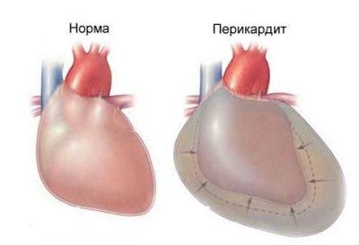
Acute pericarditis
Description of the disease
Inflammation of the pericardial sac, which is characterized by pain behind the sternum, aggravated by breathing. It can be caused by bacteria, often occurs with rheumatism, kidney disease, pneumonia, heart attack and tuberculosis.
The nature of the pain
Pain sensations can be dull or sharp, are intense, aggravated by inhalation, movement, a sudden change in posture. They can last for several days, there are heart murmurs. The patient's pulse and pressure does not change, but shortness of breath appears.
Localization
Pain on the left side of the chest, irrigating in the region of the shoulder blades.
Physician Intervention
The disease is treated by a therapist and a cardiologist.
Diagnostics
You can diagnose the disease on the basis of an ECG, blood tests for the level of aspartic aminotransferase. With the help of an x-ray, the doctor will see changes in the pericardium.
In some cases, to identify the disease, the doctor takes a puncture.
Treatment
Methods of treatment depend on the type of pathology and their nature. In the acute form of pericarditis, hospitalization should be carried out, and the chronic course of the disease can be treated on an outpatient basis.
- Diet. Rational nutrition with restriction of fats of animal origin. The patient should not drink alcohol, should reduce the amount of salt and water consumed.
- Treatment with anti-inflammatory drugs to eliminate the symptoms of the disease. Pain relief therapy is also used.
- The use of diuretics to remove fluid, antibiotics, antifungal agents.
- Stimulation of immunity.
- The operation is used when the patient has severe consequences and the inflammatory process threatens the patient's heart, for example, purulent phenomena, "shell heart".
Atrial fibrillation
 Description of the disease
Atrial fibrillation characterized by a disturbed heart attack, periods of excitations and contractions of the patient's atria and individual muscle fibers. Heart contractions increase to 600 per minute. If the attack lasts more than 2 days, then the patient develops coronary disease. Thrombus formation is possible. In a chronic course, there is a violation of the blood supply to the heart.
The nature of the pain
In some cases, the pathology runs without any symptoms, but most often a person begins to have chest pains, the heartbeat becomes more frequent. Possible interruptions in the work of the heart and stop.
The patient becomes weak, dizzy, sweating increases, shortness of breath occurs, the patient begins to choke. With constant heart disease, the muscle is depleted, congestion occurs in the internal organs, shortness of breath when climbing stairs. At night, the patient may have pulmonary edema, asthma attacks with wheezing.
The patient may lose consciousness, there are paralysis of the limbs, the pressure drops sharply, there are respiratory and cardiac arrests.
Localization
The pain is localized in the region of the heart, gives into the hands, less often in the jaw or peritoneum.
Physician Intervention
Treatment requires treatment prescribed by a cardiologist.
Diagnostics
The doctor recognizes atrial fibrillation visually by examining the patient, counting the pulse, hearing wheezing in the patient's lungs. The patient's blood pressure drops sharply during an attack.
If the doctor finds it difficult to diagnose, then prescribes:
- Holter monitoring
- Ultrasound of the heart with dopplerography.
- X-ray of the sternum
- CT scan
Treatment
- Drug treatment occurs through the appointment of thrombolytic enzymes, diuretics, beta-blockers, anticoagulants, and other medications. The course of treatment lasts several years.
- Surgical intervention. Patients are recommended breathing exercises to equalize the heart rhythm.
- Specially designed exercise therapy complex
- Operation. This method is used in cases where all other methods are ineffective. Catheter ablation is aimed at cauterization of the damaged area. Ablation with a pacemaker is characterized by transferring the work of the heart to an artificial pacemaker. The implantation of a defibrillator is used to eliminate an arrhythmia attack. Labyrinth-type surgery redirects current impulses to normalize the work of the heart.
- Folk recipes. These are compositions from natural ingredients: viburnum, yarrow, dill seeds, walnuts, honey, etc.
mitral valve prolapse syndrome
Description of the disease
 Pathology characterized by dysfunction of the left ventricular valve of the heart(growth of valve tissues, myxomatous degeneration). There are no violations in other organs. The posterior or anterior cusp of the left valve is involved in the process. Sometimes accompanied by deformation of the bone tissue of the sternum.
It can be caused by coronary heart disease or chronic rheumatism of the heart.
The nature of the pain
It is characterized by periodic pain in the middle of the chest. In the early stages, it is often asymptomatic, and is detected during a medical examination. The patient is unwell, his temperature rises to 37.5 degrees for a long period of time. Sweating increases, migraine attacks begin in the morning and evening. At night, the patient may suffocate, taking convulsive deep breaths. Pain that begins in the region of the heart is not relieved by drugs, arrhythmia begins. Doctors detect the presence of a heart murmur.
Localization
Pain is manifested in the region of the heart, giving to the middle of the chest.
Physician Intervention
To prescribe treatment, a consultation with a cardiologist and a therapist is necessary.
Diagnostics
Ultrasound, echocardiography, electrocardiography are used for diagnosing.
Treatment
- It is necessary to normalize the daily routine, sleep at least 8 hours, do not overload the body with physical activity
- A course of psychotherapy, auto-training, acupuncture, electrophoresis helps well.
- Massage of the spine
- Doctors prescribe beta-blockers, cardiotrophics, sedatives
- Possibly a course of antibiotics
Pathologies of large vessels
Aortic dissection
Description of the disease
Dissection of the aorta dangerous pathology which can result in death for the patient. It is characterized by damage to the walls of the aorta, creates an extra channel.
A complication may be bulging of the walls of the vessels in places where they become less durable.
The nature of the pain
The pain is sharp, intense, occurs suddenly, the patient's legs may go numb (usually the left one). Pain is described by patients as tearing. If with angina pectoris the pain tends to subside, then these discomfort continue both at rest and when changing positions.
The patient's blood pressure rises sharply, and then the pressure decreases, sweating becomes stronger. The pulse is asymmetrical, the person feels tired and weak. The skin becomes bluish and pale. Shortness of breath appears, the person wheezes and breathes noisily. Sometimes the bundle leads to loss of consciousness and coma.
Localization
It starts to hurt in front of the sternum (proximal bundle), pain behind the chest or behind the shoulder blade most often manifests itself with the distal nature of the pathology.
As the aorta dissects, the pain begins in the neck, jaw, between the shoulder blades, irrigates in the back and groin.
Physician Intervention
Urgent treatment by a vascular surgeon is needed, the pathology is mainly corrected only by surgery.
Diagnostics
To diagnose the disease, doctors use the following methods:
- X-ray of the sternum
- Transesophageal echocardiography
- CT angiography
- magnetic resonance angiography
- Blood tests
Treatment
The main treatment for aortic dissection is urgent hospitalization in intensive care and surgery. Doctors stop pain and bring a person out of a state of shock with analgesics or narcotic substances.
The patient's condition is stabilizing. In rare uncomplicated cases, medical treatment is possible. If other types of treatment are ineffective, then the patient is operated on by plastic surgery of the aortic valve, prosthesis, transplantation of the coronary arteries.
Pulmonary embolism
 Description of the disease
This blockage of the arteries of the lungs by a thrombus is often the result of labor activity, begins after operations, lung injuries, etc. Pathology is in second place in terms of deaths after cardiovascular diseases and oncology. The disease is difficult to diagnose, which is associated with such a high mortality rate. At the autopsy of the body of patients, statistics show that in half of them the disease was not diagnosed.
With timely treatment of the disease, the number of deaths is reduced to 10%.
The nature of the pain
The symptoms of this pathology are very diverse, which complicates the diagnosis. It all depends on the rate of development of negative processes in the lungs, the stage of the disease and the prerequisites that caused pulmonary embolism.
Pain sensations depend on the location of the thrombus, for example, if it is in a large artery, then the patient may experience only shortness of breath and nothing more, and when small vessels overlap, the pain is sharp, burning, intense.
A person develops shortness of breath. A strong heartbeat begins, the patient's skin turns pale, becomes gray, the intestines are disturbed, tension arises in the abdominal region. The aorta pulsates, heart murmurs begin, pressure drops.
The veins of the neck and abdominal region fill with blood, begin to swell.
Localization
Pain is localized in the middle of the chest, resembling signs of pleurisy.
Physician Intervention
Treatment requires the intervention of a vascular surgeon.
Diagnostics
The following methods are used for diagnosis:
- x-ray
- CT scan
- echocardiography
- Ultrasound examination of veins
- Scintigraphy
Treatment
- Drug treatment: intravenous fraxiparin, sodium heparin; oral warfarin;
- Use of drugs to dissolve blood clots, eg streptokinase, urokinase, alteplase.
- Doctors can apply the following measures: indirect heart massage, oxygen therapy, artificial ventilation of the lungs, the introduction of saline solutions with droppers.
- Operation. This may be the elimination of an embolus or an artery wall with a plaque. Often, plastic surgery of the heart is necessary, due to stretching of the right ventricle.
- Installing a cava filter, which allows you to prevent the disease.
Respiratory diseases
Laryngotracheitis, bronchitis
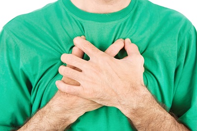 Description of the disease
This inflammatory diseases developing as a result of infection, accompanied by cough, chills, respiratory complications.
The nature of the pain
Pain can be sharp or dull, but will certainly increase with coughing. The patient complains of a sore throat, he has a soreness behind the sternum, there is an excruciating cough, in which pain is felt.
There may be an increase in lymph nodes in the neck.
Localization
The pain begins behind the chest when coughing.
Physician Intervention
This disease is treated by a pediatrician, pulpologist, otolaryngologist.
Diagnostics
The doctor detects the disease on the basis of a visual examination of the patient, with complaints of the patient, while listening. If necessary, X-rays, computed tomography, MRI are done.
Blood tests, sputum smears are carried out. Sometimes a biopsy of the mucous membrane of the larynx is done.
Treatment
- Gentle mode for the vocal cords
- Diet with no pickles, hot or cold foods, hot spices. Supplemented with abundant alkaline drink.
- Drug treatment with antiviral drugs (cyclofen, Ingavirin); antibiotics (Flemoklav, Azithromycin); antihistamines (Zirtek, Suprastin); drugs that relieve inflammation (Paracetamol).
- Be sure to use cough medicines (Sinekod, Codelac) and expectorants, mulokilitiki (Mukaltin, Dr. Mom).
- The use of homeopathic remedies.
Pleurisy
Description of the disease
Inflammation of the pleura (the membrane that protects the lungs) due to bacteria (streptococci, fungi, pneumococci, etc.), trauma, tuberculosis, allergic reactions.
The nature of the pain
Pain is often can be confused with heart, doctors rule out the possibility of a heart attack ECG. When a person lies on the affected area, the discomfort decreases. The nature of the pain is intense, the patient has a fever, it is difficult for him to breathe, coughing also occurs with pain.
The patient's pulse becomes frequent.
Localization
Usually pains are localized behind a breastbone, become stronger at an inhalation-exhalation. With atypical forms, irrigation into the abdomen is possible, so sometimes pleurisy can be confused with an attack of appendicitis.
Physician Intervention
The disease is treated by a pulpologist.
Diagnostics
- To diagnose the disease, an x-ray is needed to detect blackouts, accumulations of fluid.
- Puncture of fluid in the pleura
- Biochemistry of blood
- Pleura biopsy
- Patient examination
- Listening to the lungs
- Palpation of the chest
Treatment
- The patient must comply with bed rest with wiping with a damp cloth soaked in warm water.
- Removal of inflammation with the help of hormonal, antibacterial, decongestant drugs.
- Symptomatic treatment: lowering the temperature with antipyretics, pain relief with analgesics, increasing the body's defenses with immunostimulants.
- Physiotherapy with heating of the sternum
- Pumping out the contents of the pleura, which does not eliminate the compression of internal organs
- Local treatment: rubbing rubs, applying compresses and bandages.
- Recovery with the help of exercise therapy, yoga, taking vitamins and minerals, good nutrition.
Cancer (lung, bronchi, pleura, lymph node metastases)
 Description of the disease
A malignant neoplasm of the lung caused by inhalation of carcinogens, smoking, harmful production.
The nature of the pain
In the early stages, the disease is asymptomatic, it just looks like bronchitis in the chronic stage or ARVI disease. Therefore, cancer is often diagnosed at an advanced stage of the disease. With the central form of oncology, the pathology manifests itself faster due to respiratory disorders.
First, there is a dull pain in the chest, which occurs periodically; then the pain becomes so strong that the person cannot sleep, only narcotic substances alleviate the condition. Cough is often accompanied by sputum with blood, visually similar to pink or red jelly. Shortness of breath begins.
The patient is dizzy, the temperature rises, weight loss occurs.
Localization
Pain is localized in the chest at the site of the focus of the disease, but when the tumor grows, it can begin in the area of \u200b\u200bthe ribs, in the region of nearby internal organs.
Physician Intervention
The disease is treated by an oncologist.
Diagnostics
Cancer is diagnosed by the following methods:
- X-ray in two projections
- Fluorography
- Bronchoscopy
- Mediascopy
- Biopsy of lymph nodes, internal organs
Treatment
- Surgical intervention
- Chemotherapy aimed at suppressing the growth of tumor cells. It is done with the help of Vincristine, Carboplatin, Irinotecan, Etoposide.
- course of radiation therapy. Basic methods: technology (IGRT) (irradiates the affected cells, corrects, transfers the load to another part of the tissue); brachytherapy (moving to the cells of means that enhance their action); "smart knife" (the exact impact of the cyber-knife on the affected cells).
- Hormonal correction
- Immunotherapy
Diseases of the gastrointestinal tract
Gastroesophageal reflux disease (GERD)
 Description of the disease
Chronic pathology of the gastrointestinal tract that occurs due to periodic reflux of food into the esophagus with subsequent damage to the mucous membranes, nearby organs (bronchi, pharynx, trachea).
The nature of the pain
The patient complains of heartburn, sour belching. Often pain burning sensations begin when swallowing and after eating, in rare cases vomiting occurs, followed by hiccups. The patient may complain of pain in the chest, resembling a heart attack or angina pectoris.
At night, due to throwing into the esophagus, the patient begins to cough, his throat tickles, hoarseness occurs, bronchitis or asthma may develop.
Symptoms are aggravated by physical exertion, after eating.
Localization
Burning pain, accompanied by heartburn, is localized in the epigastric region, irrigates in the cervical region, shoulders. Starts 1-2 hours after eating. Can become stronger after drinking soda, when moving.
Physician Intervention
For diagnosis and treatment, consultation with a general practitioner and a gastroenterologist is necessary.
Diagnostics
- Proton pump inhibitor test.
- Intra-food pH monitoring
- Fibroesophagogastroduodenoscopy
- Chromoendoscopy of the esophagus.
- Ultrasound of internal organs
- X-ray.
- Blood and stool tests
Treatment
- Surgery is needed only in extreme cases.
- Compliance with diet and special recommendations. Hot dishes are excluded from the diet, you can not eat a lot of food for dinner, eat citrus fruits, chocolate and fat. You can not make sharp bends forward after eating, it is not recommended to wear tight clothes and corsets so as not to raise intra-abdominal pressure.
- Weight loss is recommended in the presence of obesity.
- You can not drink antispasmodics, prostaglandins.
- A course of medications: antacids (Almagel), prokinetics (Motilium).
Esophagitis
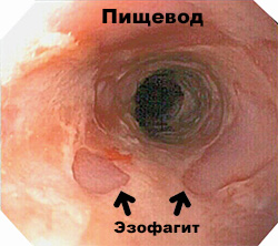 Description of the disease
This is a pathology characterized by inflammation of the mucous membranes. If the disease is not treated, then the complications will be a peptic ulcer, the occurrence of perforation of the esophagus. There are different types of pathology: the disease is both acute and chronic. It affects the superficial layers of the mucosa or deep.
The lesion of the esophagus can be complete, in some cases, the inflammation extends only to the upper or lower section.
The nature of the pain
Manifestations of pain depend on the inflammatory process. For example, in the catarrhal form, the esophagus is sensitive only to cold and hot dishes. In severe forms, intense burning pains appear. Swallowing also causes pain. The patient complains of increased salivation, heartburn, vomiting with blood is possible. Then the symptoms disappear, but within 3 months, scars appear on the walls of the esophagus.
Localization
Soreness is felt behind the sternum, can be irrigated in the cervical region and in the back.
Physician Intervention
For the treatment of the disease, it is necessary to consult a surgeon and a gastroenterologist.
Diagnostics
- x-ray
- esophagoscopy,
- endoscopic mucosal biopsy
- Histological examination
- Esophagomanometry
Treatment
- Medications: antacids and drugs from the famotidine group.
- Diet with the exception of alcoholic beverages, food that increases the secretion of gastric juice
- Course of antibiotics
- Refusal of tranquilizers and sedatives
- Physiotherapy (electrophoresis, mud therapy).
- Operation. It is done in extremely severe cases with scarring of the tissues of the esophagus.
Foreign bodies of the esophagus
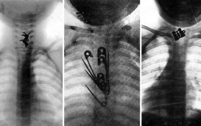 Description of the disease
Foreign bodies in the esophagus don't happen that often, but getting coins, bones, prostheses, food, small parts of toys into it requires quick help. Objects can get stuck in it due to the large volume of the object in comparison with the lumen of the esophagus, tumors, etc.
The nature of the pain
If toys do not have sharp edges, then parents often do not suspect that the lost object is stuck in the esophagus. When a body with sharp edges enters the esophagus, strong salivation is noticed. Such children refuse food, do not want to turn their heads once again. When hit by bodies with a smooth surface, the patient may cough, drink, but do not eat thick food.
Respiratory disturbances are observed.
The patient feels thirsty, loses weight from lack of water. When an object is in the upper part of the esophagus, the larynx moves forward and becomes wider. If palpation is carried out in front of the larynx, then aching pain occurs, which disappears as soon as the doctor removes the fingers. The patient is shivering, there may be a temperature
Localization
Pain develops below the place where the foreign body is
Physician Intervention
For consultation and diagnosis, the help of a surgeon and a therapist may be needed.
Diagnostics
- Visual examination of the patient
- Esophagoscopy
- x-ray
- Fistulography
Treatment
The main action that the doctor can take is to remove the object. This can be done in a gentle way with the help of enveloping substances, a special diet, washing with furacillin.
If the foreign body is not removed, then an endoscopy with extraction under general or local anesthesia should be performed.
Stomach ulcer
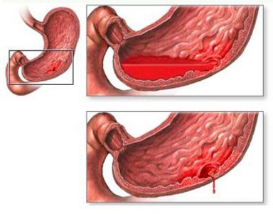 Description of the disease
This is a chronic disease with frequent relapses. With it, defects are formed in the stomach with penetration under the mucous membranes. During the disease, periods of remission and exacerbation alternate.
The nature of the pain
The clinical picture of the pathology can be different, it depends on the pain threshold of the patient, the stage of the disease, the location of the focus, and the individual characteristics of the person. The nature of the pain is constant and periodic (at night, after eating). The soreness observed in the patient is pulling, dull, acute, etc. Its intensity also varies: from severe to slightly noticeable soreness.
The patient complains of heaviness in the abdomen after eating, belching, heartburn, decreased appetite and delayed defecation.
Localization
Pain is observed in the left side of the chest, radiates to the arm or dorsal region, ribs, lower back, pelvic region.
Physician Intervention
For treatment, it is necessary to consult a surgeon and a gastroenterologist.
Diagnostics
Identification of the disease is carried out using a blood test, fibroesophagogastroduodenoscopy, X-ray, pH-metry and daily monitoring of the pH of gastric juice.
Treatment
- A course of antibiotics (Amoxicillin, Tetracycline).
- Treatment with antisecretory drugs that neutralize hydrochloric acid(Maalox).
- Taking Ranitidine, Omeprazole, Sucralfate, De-nol, etc.
- It is recommended to take antidepressants and tranquilizers.
- To relieve pain, antispasmodics are used, for example, no-shpa.
- Prescribe treatment with probiotics, for example, Lineks.
Be sure to watch the following video
In which case immediately see a doctor?
- With a sharp attack of pain, especially if the patient suffocates, weakens, his eyes darken.
- If Nitroglycerin Tablets Cannot Relieve Pain
- With loss of consciousness.
First aid
First aid depends on the disease:
- With angina pectoris, they give a nitroglycerin tablet, if it does not help, they call an ambulance.
- In case of a heart attack, they provide the maximum flow of oxygen to the patient and call an ambulance.
- To relieve spasm in osteochondrosis, it is enough to take a pill that relieves pain or inflammation, make local rubbing with non-steroidal ointment.
- With an ulcer, medical attention is urgently needed.
- With pain from severe heartburn, Peptobismol is taken.
- In case of injury, the patient is placed on the affected side and the chest is fixed. Urgent transportation of the patient to the traumatologist is necessary.
- Neuralgia is relieved by warm or cold compresses.
- In case of a sharp pain attack with loss of consciousness, it is necessary to check whether the person is breathing, whether his heart is beating, to do cardiopulmonary resuscitation. It is laid so that the legs are higher than the head. The patient must be tried to bring to himself and call a doctor.
Drawing conclusions from the foregoing, one can understand that pain in the sternum in the middle does not appear just like that - more often it is a symptom of a serious illness.
Therefore, you do not need to let everything take its course, doing self-treatment. It is necessary to consult a doctor for timely diagnosis of the disease and its treatment. This the right way prevent an attack of angina pectoris, heart attack, get rid of stomach ulcers and other serious pathologies that poison the existence.
In contact with